Glycogen is a branched homopolymer composed of α-D-glucose units serving as a storage form of the monosaccharide, and therefore of energy.[1]
As it is synthesized without a template, unlike proteins and nucleic acids, it exists as a population of molecules of varying structures and sizes.[2][3]
Glycogen is a compact and soluble macromolecule exerting low osmotic pressure and allowing rapid glucose release when needed.[4]
It forms assemblages with proteins essential for its metabolism and anchoring to the cytoskeleton and membranes. Such aggregates are called β-granules. Furthermore, phosphate groups are covalently linked to the polysaccharide.[5][6]
In animals, glycogen is found in practically all cells and, in mammals, it is most abundant in the liver and skeletal muscle. Within the liver, several β-granules assemble to form the so-called α-granules.[3] Glycogen is also found in lysosomes.[7]
In humans, it represents less than 1% of the body’s energy stores and is essential for maintaining blood glucose homeostasis.[8]
It is absent in plants, where starch serves as the storage form of glucose.[9]
Glycogen has little nutritional significance for humans.[10]
Summary: Key Points
- Definition and structure: glycogen is a branched homopolysaccharide of α-D-glucose with α-(1→4) linear bonds and α-(1→6) branch points every 8–12 residues, bound to glycogenin.
- Enzymatic regulation: controlled via allosteric effectors (AMP, ATP, glucose 6-phosphate, Ca2+) and covalent modifications triggered by insulin, glucagon, and epinephrine.
- Degradative pathways: degraded in the cytosol by glycogen phosphorylase and debranching enzyme, and in lysosomes by acid α-glucosidase.
- Tissue distribution and storage: stored in the liver to maintain euglycemia, and in skeletal muscle for local ATP production.
- Energetic efficiency: highly efficient storage form, conserving ≈ 96% of potential energy under aerobic conditions and ≈ 60% under anaerobic oxidation.
Contents
- Historical background
- Chemical and molecular structure of glycogen
- β-Granules
- α-Granules
- Glycogen metabolism: the synthetic phase
- Glycogen metabolism: the degradative phase
- Coordinated regulation of glycogen metabolism
- Localization of glycogen in humans
- Why it is important to humans
- Glycogen and muscle work
- Energy yield under anaerobic conditions
- Energy yield under aerobic conditions
- References
Historical background
Glycogen was discovered in 1857 by the French physiologist Claude Bernard, who is considered the founder of experimental medicine.[11] In the second half of the 20th century, studies on glycogen metabolism led to several significant discoveries such as:
- the reversible phosphorylation of proteins;
- protein kinases and protein phosphatases;
- the effect of insulin on the activity of intracellular enzymes.[9]
These landmark discoveries led to the award of four Nobel Prizes, three for Physiology or Medicine, to Carl Ferdinand Cori and Gerty Theresa Cori (née Radnitz) in 1947, Earl Sutherland Jr. in 1971, and Edwin Krebs and Edmond Fischer in 1992, and one for Chemistry, to Louis Leloir in 1970.[12]
Chemical and molecular structure of glycogen
An individual glycogen molecule is a branched polymer of α-D-glucose in pyranose form, that is, a rigid six-membered heterocyclic ring composed of five carbon atoms and one oxygen atom, with a chair conformation.
The central priming protein glycogenin (EC 2.4.1.186) and phosphate groups are covalently bound to the polysaccharide chain.[1]
Most of the glucose units are linked by α-(1→4)-glycosidic bonds, where each unit is connected to the next through a bond between the C-1 atom of one unit and the hydroxyl group on the C-4 atom of the next, with an oxygen atom acting as a bridge between the two carbons.
Branch points are introduced by α-(1→6)-glycosidic bonds, which occur approximately every 8–12 residues, again with an oxygen atom bridging the two carbons (C-1 and C-6), and with an average chain length of about 13 residues in mammals. Because each branch terminates in a non-reducing residue, there are n + 1 non-reducing ends in the molecule, where n is the number of chains, but only one reducing end, to which glycogenin is linked.[13]
Note that in in disaccharides, oligosaccharides, and polysaccharides, the non-reducing end is the end that lacks a free anomeric carbon atom.
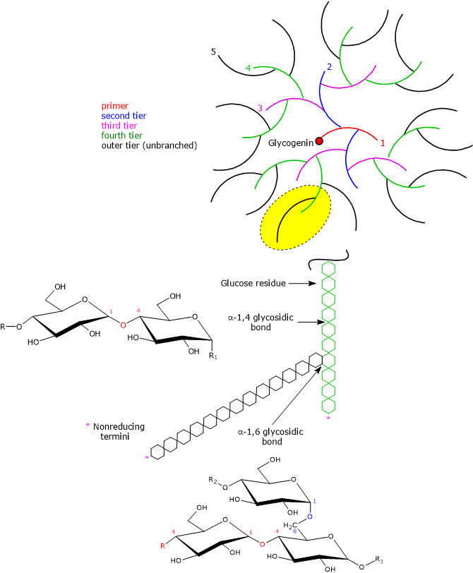
Because it features the same types of bonds, the primary structure of glycogen resembles that of amylopectin, which, together with amylose, is one of the two polymers of D-glucose units composing starch. However, compared with amylopectin, where branches occur every 25–30 glucose units, glycogen is more highly branched, and the branches are shorter.[14]
Heterogeneity of glycogen structure and particle size
Unlike proteins and nucleic acids, polysaccharides are synthesized without a template, through the addition of monosaccharides or oligosaccharides to the growing chain. Furthermore, because branching occurs without precise localization, molecules with the same mass do not necessarily share the same structure. Hence, for each type of molecule, there are multiple chemical structures.[3]
Moreover, glycogen isolated from different biological sources exists as a population of molecules with different sizes. Therefore, the best way to describe its chemistry is to define the distribution of molecular masses, as well as the average frequency and length of branches.[2]
Finally, it should be emphasized that glycogen is not a static entity but constantly varies over the course of its existence.[5]
As glucose has chiral centers, it exists as a pair of enantiomers, designated according to the Fischer–Rosanoff convention as D-glucose, the most widespread in nature and the monomeric unit of glycogen and starch, and L-glucose.[15]
Factors stabilizing the 3D structure
The folding of macromolecules such as proteins, nucleic acids, and polysaccharides into three-dimensional structures is governed by the same universal principles. Their monomeric units, namely amino acids, nucleotides, and monosaccharides, each with varying structural rigidity, are joined by covalent bonds to form linear polymers that spontaneously fold into three-dimensional conformations stabilized by noncovalent interactions, such as:
- hydrogen bonds,
- van der Waals interactions,
- hydrophobic interactions, and
- ionic interactions, when charged subunits are present.[13]
These interactions can occur within a single macromolecule or between different macromolecules, as seen in supramolecular complexes such as cellulose or multienzyme complexes.[16]
Because the pyranose ring of glucose is a rigid structure, the three-dimensional conformation of oligosaccharides and polysaccharides results from rotation about both C–O bonds of the glycosidic linkage, with the dihedral angles denoted as Φ (phi) and Ψ (psi). However, rotation about each C–O bond is restricted due to steric interference from substituents. Consequently, certain conformations are energetically more stable than others. For amylose and glycogen, the most stable 3D structure is a tightly coiled helix stabilized by interchain hydrogen bonds.[9][17]
Advantages of the branched structure
The highly branched structure of glycogen offers several major physiological advantages:
- The non-reducing ends exposed on the outermost tier act as substrates for glycogen phosphorylase (EC 2.4.1.1). Consequently, multiple glycogen phosphorylase molecules can work simultaneously, enabling rapid mobilization of stored glucose in the form of glucose 1-phosphate (G1P).[14]
- Branching allows stored glucose to exert a dramatically lower osmotic pressure than it would in monomeric form. For example, hepatocytes store an amount of glucose that, if free, would correspond to a concentration of approximately 0.4 M, compared with a glycogen particle concentration of only about 0.01 mM. If glucose were free in the cytosol, the resulting high osmolarity would cause an osmotic influx of water leading to cell lysis. Furthermore, given an extracellular glucose concentration of ≈ 5 mM, glucose uptake against an intracellular gradient of 0.4 M would be energetically prohibitive.[9]
- Extensive branching allows the formation of dense, compact granules.
- If branches were absent or scarce, an excessively large number of long linear polymers would be required to yield a comparable number of non-reducing ends and store equivalent amounts of glucose, likely causing structural cell damage. Evidence for this comes from Andersen’s disease (amylopectinosis or glycogen storage disease type IV), a rare genetic disorder caused by mutations in the branching enzyme gene; the resulting enzyme deficiency leads to the tissue accumulation of poorly branched, insoluble glycogen resembling amylopectin.[2][6]
- Finally, branching maintains the macromolecule in a soluble state, unlike linear starch components.[5]
Whelan’s model
Due to the sequential action of glycogenin, glycogen synthase (EC 2.4.1.11), and the branching enzyme, also known as glycosyl-(4,6)-transferase (EC 2.4.1.18), the glycogen molecule expands exponentially in concentric tiers around the central glycogenin core. According to Whelan’s structural model, two distinct types of glucose chains are present:
- A-chains, which are unbranched and located exclusively on the outer surface;
- B-chains, which are internal and each contain, on average, two branch points.[18][19]
Theoretical calculations indicate a maximum physical limit of 12 tiers, corresponding to a diameter of approximately 42 nm, ≈ 55,000 glucose units, and a molecular mass of around 107 Da.[20][21]
Assuming a spherical shape with each tier contributing 3.8 nm in thickness, progression from the third to the twelfth tier results in:
- a 5.4-fold increase in diameter;
- a 156-fold increase in volume (proportional to the cube of the radius);
- a 45.6-fold increase in carbohydrate content; and
- an exponential increase in the number of A-chains in the outermost tier, equal to 2n − 1, where n represents the tier number.[18]
| Level | Diameter (nm) | Chains/Level | Glucose/Level | Total Glucose |
|---|---|---|---|---|
| 1 | — | 1 | 13 | 13 |
| 2 | 3.8 | 3 | 26 | 39 |
| 3 | 7.8 | 7 | 52 | 91 |
| 4 | 11.6 | 15 | 104 | 195 |
| 5 | 15.4 | 31 | 208 | 403 |
| 6 | 19.2 | 63 | 416 | 819 |
| 7 | 23.0 | 127 | 832 | 1,651 |
| 8 | 26.8 | 255 | 1,664 | 3,315 |
| 9 | 30.6 | 511 | 3,328 | 6,643 |
| 10 | 34.4 | 1,023 | 6,656 | 13,299 |
| 11 | 38.2 | 2,047 | 13,312 | 26,611 |
| 12 | 42.0 | 4,095 | 26,624 | 53,235 |
In skeletal muscle, electron microscopy analysis of glycogen particle size reveals predominantly sub-maximal particles with an average diameter of ≈ 25 nm, corresponding to seven tiers.[22]
Features of Whelan’s model
An important feature of Whelan’s model is that the outermost tier contains, in the form of A-chains, approximately 50% of all glucose residues. However, not all of these residues are immediately accessible to glycogen phosphorylase, as the enzyme stops four glucose residues short of a branch point. The subsequent intervention of the glycogen debranching enzyme (EC 2.4.1.25 and EC 3.2.1.33), whose catalytic rate is slower than that of glycogen phosphorylase, removes the branch, allowing glycogenolysis to proceed.[2][3]
Why the 13th tier is not possible
Formation of a 13th tier is structurally impossible due to severe steric hindrance arising from the high density of glucose units on the particle’s surface. This overcrowding restricts the physical access of catalytic sites of metabolic enzymes, including glycogen synthase, to the growing outer chains.[2][23]
Furthermore, mathematical modeling demonstrates that the physiological parameters of mammalian glycogen, namely a branch length of ≈ 13 residues, an average branching frequency of 2 per tier, and a maximum limit of 12 tiers, are precisely optimized to maximize the number of glucose molecules available for rapid mobilization per unit time.[24]
Glycogenin
The glycogen macromolecule incorporates the self-glucosylation protein glycogenin, which remains covalently bound to the reducing end of the core polysaccharide chain. Glycogenin initiates glycogen synthesis via autoglycosylation, catalyzing the attachment of 7–11 glucose units to a specific tyrosine residue (Tyr194). This short primer then serves as the substrate for glycogen synthase. Additionally, by binding to actin filaments, glycogenin anchors the nascent glycogen granule to the cytoskeleton.[14][25]
Phosphate groups
In addition to glycogenin, the glycogen macromolecule contains low levels of covalently bound phosphate groups.
For decades, these phosphate residues were considered experimental artifacts or contaminants whose abundance inversely correlated with sample purity. It was not until the early 1980s that they were recognized as integral components of the polysaccharide, covalently linked to C-2 and C-3 positions as monoesters, likely arising from rare catalytic side reactions of glycogen synthase.[26]
Accumulating evidence indicates that glycogen phosphorylation plays a physiological role in granule turnover, analogous to its role in plant starch metabolism. Supporting this view is the identification of laforin, a dual-specificity glycogen phosphatase; loss-of-function mutations in the laforin gene cause Lafora disease, a fatal progressive myoclonus epilepsy characterized by the accumulation of hyperphosphorylated, insoluble glycogen-like aggregates (Lafora bodies).[27]
The exact physiological mechanisms governed by phosphate groups remain under investigation, with two main hypotheses proposed:
- Phosphate monoesters might disrupt internal hydrogen-bonding networks, thereby exposing hydrophobic regions and driving self-aggregation and insolubility. Dephosphorylation by laforin maintains structural solubility, ensuring continued enzymatic branching and elongation.[28]
- Alternatively, progressive accumulation of phosphate monoesters may track the molecular age of the glycogen granule, serving as a quality-control signal. Hyperphosphorylation-induced loss of solubility could act as a metabolic marker that targets older granules for selective lysosomal degradation via macroautophagy (glycophagy) rather than cytosolic glycogenolysis.[29]
β-Granules
Individual glycogen molecules are too small to be resolved by light microscopy. However, transmission electron microscopy has identified three distinct levels of glycogen organization: β-granules, γ-particles, and α-granules.[2]
β-Granules, present in the cytosol of bacteria, Archaea, fungi, and animal cells, comprise the glycogen polysaccharide, the initiator protein glycogenin, and γ-particles (protein-rich structures ≈ 3 nm in diameter). β-Granules have an estimated molecular mass of ≈ 108 Da, a diameter of 20–30 nm, and display a characteristic rosette-like appearance under electron microscopy. Functionally, they serve as a rapidly accessible energy reserve.[5][14]
Under physiological conditions, associated proteins constitute a major portion of the granule’s total mass. These proteins interact with one another, the cytoskeleton, or organelle membranes, and coordinate glycogen metabolism. Key protein components include:
- enzymes directly catalyzing turnover: glycogen synthase, the glycogen debranching enzyme, and glycogen phosphorylase;
- regulatory and scaffolding proteins, such as:
- laforin and phosphoprotein phosphatase 1 (PP1; EC 3.1.3.17);
- phosphorylase kinase (EC 2.7.11.19) and AMP-activated protein kinase (AMPK; EC 2.7.11.31);
- membrane-anchoring proteins such as Stbd1 (starch-binding domain-containing protein 1);
- malin, an E3 ubiquitin ligase (EC 2.3.2.27) that targets glycogen-associated proteins via interactions with laforin and TRIM7.[30]
Unlike static macromolecular assemblies such as the pyruvate dehydrogenase complex or ribosomes, the protein stoichiometry and composition of β-granules are highly dynamic, adjusting rapidly via association-dissociation events in response to metabolic signals. Furthermore, structural and compositional heterogeneity exists across different tissues and even within distinct subcellular niches of the same cell, such as skeletal muscle.[3]
α-Granules
In hepatocytes, individual β-granules assemble into higher-order supramolecular structures termed α-granules.
Composed of clusters of multiple β-granules, α-granules possess a molecular mass exceeding 108 kDa and can reach diameters up to ≈ 300 nm, exhibiting a broccoli-like morphology under electron microscopy. α-Granules are mobilized more slowly than β-granules, providing a sustained glucose supply.[31]
Although the precise mechanism of α-granule assembly remains under active investigation, current models suggest that β-granules are covalently linked through a protein scaffold rich in disulfide bonds.[3]
Glycogen metabolism: the synthetic phase
Glycogen accumulation occurs during periods of energy surplus. Glycogenesis takes place in the cytosol in association with actin filaments, utilizing glucose derived either from dietary carbohydrates or synthesized de novo from non-carbohydrate precursors (such as lactate and alanine).[3][32]
Lactate, generated by active skeletal muscle during anaerobic exertion, by erythrocytes via continuous glycolysis, and by other tissues, is transported to the liver, where it serves as a primary substrate for gluconeogenesis. The newly synthesized glucose is released into the circulation and taken up by skeletal muscle, where it can be stored as glycogen or re-converted to lactate, completing the Cori cycle.[33]
Alanine, produced in extrahepatic tissues via transamination of glycolytic pyruvate, acts as a dual carrier of carbon skeletons and amino nitrogen. Transported to the liver, its carbon skeleton fuels gluconeogenesis while its amino group is detoxified via the urea cycle. Glucose released into the bloodstream returns to peripheral tissues, where it can be converted back to pyruvate, completing the glucose-alanine cycle.[34]
Glucose entry into target cells is mediated by facilitated diffusion via glucose transporter proteins (GLUTs). Notably, GLUT4 is the principal insulin-responsive transporter, primarily expressed in skeletal muscle, cardiac muscle, and adipose tissue.[13]
Glucose uptake and phosphorylation
Upon entry into the cytosol, glucose is rapidly phosphorylated to glucose 6-phosphate (G6P). This reaction is catalyzed by glucokinase (hexokinase IV; EC 2.7.1.1) in hepatocytes and pancreatic β-cells, and by hexokinases I–III in other cell types.[35]
Fate of glucose 6-phosphate
Glucose 6-phosphate sits at a major metabolic branch point. Depending on the cellular energy state and hormonal signals, it can enter the glycolytic pathway to produce energy and/or precursors for other metabolic pathways. Alternatively, it can undergo isomerization to glucose 1-phosphate, an example of positional isomerism, in the reversible reaction catalyzed by phosphoglucomutase (EC 5.4.2.2). While G1P is used for glycogen synthesis, glucose 6-phosphate enters the pentose phosphate pathway if the cell requires NADPH for reductive biosyntheses, such as those of fatty acids or cholesterol, or ribose 5-phosphate for nucleotide synthesis.[9][36]
Glycogen synthesis
When glycogen synthesis is activated, glucose 1-phosphate is converted to UDP-glucose by UDP-glucose pyrophosphorylase (EC 2.7.7.9) in an exergonic reaction coupled to pyrophosphate hydrolysis. UDP-glucose serves as the activated glucosyl donor. Initially, glycogenin self-catalyzes the attachment of the first glucosyl residue to Tyr194, followed by autoglycosylation of 6–10 additional glucose residues to build a short oligosaccharide primer.[37] This primer then serves as the substrate for glycogen synthase, which works concertedly with the branching enzyme to drive full-scale glycogen macroassembly.[8]
Glycogen metabolism: the degradative phase
Unlike glycogen synthesis, the catabolic phase of glycogen metabolism occurs in both the cytosol and lysosomes via distinct pathway mechanisms. Cytosolic glycogenolysis is spatially linked to the endoplasmic and sarcoplasmic reticulum, mobilizing glucose to meet cellular and systemic energy demands.[38][39]
The concerted action of glycogen phosphorylase and the bifunctional glycogen debranching enzyme, exhibiting oligo-α-(1,4)→α-(1,4)-glucan transferase and amylo-α-(1,6)-glucosidase activities, yields glucose 1-phosphate (≈ 90% of total released glucose) and free glucose (≈ 10%), respectively.[14]
In skeletal muscle, high hexokinase activity rapidly phosphorylates any free glucose to G6P, trapping it within the cell for local ATP production via glycolysis. Conversely, in gluconeogenic tissues such as hepatocytes, renal cortex cells, and enterocytes, lumenal glucose 6-phosphatase (EC 3.1.3.9) in the endoplasmic reticulum catalyzes the dephosphorylation of G6P (derived from G1P isomerization) to free glucose, which exits via GLUT transporters to maintain blood glucose homeostasis.[40]
Lysosomal degradation
Although glycogenesis occurs in the cytosol, a distinct pool of glycogen is compartmentalized within lysosomes. Lysosomal glycogen uptake is mediated by an autophagic pathway (glycophagy), accounting for approximately 10% of total liver glycogen and 5% of muscle glycogen content.[41]
Progressive glycogen phosphorylation is hypothesized to regulate its lysosomal degradation. Accumulation of phosphate monoesters over time correlates with granule age and functions as a quality-control mark: hyperphosphorylation decreases granule solubility, marking older particles for autophagic sequestration and lysosomal breakdown via glycophagy.[5][42]
Within lysosomes, glycogen breakdown is catalyzed by acid α-glucosidase (GAA; EC 3.2.1.20), releasing D-glucose. Because GAA preferentially hydrolyzes α-(1→4) glycosidic linkages, the precise mechanism of α-(1→6) branch cleavage inside the lysosomal compartment remains under investigation.[43]
The physiological necessity of lysosomal glycogen turnover is highlighted by Pompe disease (glycogen storage disease type II), an autosomal recessive disorder caused by GAA deficiency. Loss of functional acid α-glucosidase leads to massive accumulation of unbranched glycogen in lysosomes, causing severe neuromuscular damage that is fatal in infancy if untreated.[7]
Coordinated regulation of glycogen metabolism
Both glycogenesis and glycogenolysis are thermodynamically exergonic; if active simultaneously within the same cellular compartment, they would create a futile cycle resulting in net ATP dissipation. Consequently, these pathways are tightly and reciprocally regulated so that activation of one pathway coincides with inhibition of the other. Evolutionarily, reciprocal regulation is achieved by utilizing distinct key enzymes for synthetic and degradative steps, analogous to the regulation governing glycolysis and gluconeogenesis.[13]
The primary regulatory hubs are glycogen phosphorylase and glycogen synthase, whose activities are fine-tuned via two main mechanisms:
- Allosteric regulation: operates on a millisecond timescale and is instantly reversible. Allosteric effectors include Ca2+ ions, free glucose, and energy-charge indicators such as ATP, AMP, and G6P.
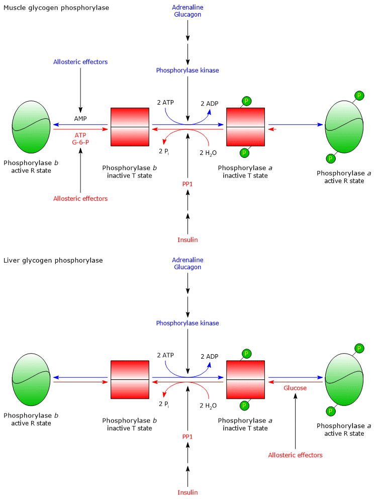
- Covalent modification: involves reversible phosphorylation and dephosphorylation of target enzymes, including glycogen phosphorylase, glycogen synthase, phosphorylase kinase, protein phosphatase 1 (PP1), and glycogen synthase kinase 3 (GSK3; EC 2.7.11.26). Covalent control occurs on a timescale of seconds to minutes and is governed by endocrine signaling, primarily insulin, glucagon, and epinephrine (adrenaline), binding to specific cell-surface receptors.[14]
These allosteric and covalent mechanisms act cooperatively to maintain precise control over cell and tissue carbohydrate metabolism.[15]
| Enzyme | Primary pathway | Active form | Covalent modification (hormonal) | Allosteric activators | Allosteric inhibitors |
|---|---|---|---|---|---|
| Glycogen synthase | Glycogenesis | Dephosphorylated (a) | Activated by insulin (dephosphorylation) Inhibited by glucagon and epinephrine (phosphorylation) |
G6P | ATP, AMP, inorganic phosphate |
| Glycogen phosphorylase | Glycogenolysis | Phosphorylated (a) | Activated by glucagon and epinephrine (phosphorylation) Inhibited by insulin (dephosphorylation) |
AMP, Ca2+ (muscle) | G6P, ATP, Glucose (liver) |
Covalent regulation
The metabolic shifts triggered by insulin, glucagon, and epinephrine binding to their plasma membrane receptors in hepatocytes and myocytes are detailed below.
Insulin
Insulin is secreted by pancreatic β-cells in response to elevated blood glucose levels (e.g., in the postprandial state) and exerts broad anabolic effects. Upon binding to cell-surface insulin receptors on hepatocytes and myocytes, it initiates a signaling cascade that promotes the net dephosphorylation of key metabolic enzymes:
- glycogen phosphorylase is dephosphorylated and inactivated;
- glycogen synthase is dephosphorylated and activated.
Simultaneously, insulin promotes the translocation of GLUT4 glucose transporters to the plasma membrane of skeletal muscle cells and adipocytes. Consequently, glycogenesis is stimulated while glycogenolysis is suppressed, driving systemic glucose clearance.[44]
Glucagon
Glucagon is secreted by pancreatic α-cells during hypoglycemia and acts as a catabolic hormone. Upon binding to G protein-coupled receptors on hepatocytes, glucagon triggers a cAMP-dependent phosphorylation cascade that results in the phosphorylation of:
- glycogen phosphorylase, converting it to its active form;
- glycogen synthase, converting it to its inactive form.
Thus, hepatic glycogen synthesis is inhibited while glycogen breakdown is activated, releasing glucose into the bloodstream to restore glycemic homeostasis.[9]
Adrenaline
Epinephrine (adrenaline) is secreted by the adrenal medulla during sympathetic nervous system activation (the fight-or-flight response) and intense exercise. Like glucagon, it exerts catabolic effects via protein phosphorylation, activating glycogen phosphorylase and inhibiting glycogen synthase.
Unlike glucagon (whose receptors are primarily restricted to hepatocytes), epinephrine acts on both liver and skeletal muscle. It binds to α1– and β2-adrenergic receptors in hepatocytes, and to β2-adrenergic receptors in myocytes.[15]
Vasopressin
Vasopressin, along with epinephrine acting via α1-adrenergic receptors, activates the phosphatidylinositol signaling pathway, triggering calcium release from the endoplasmic reticulum. Elevated cytosolic calcium levels stimulate glycogenolysis while inhibiting glycogen synthesis.[8]
Allosteric regulation
In addition to hormonal covalent control, glycogen metabolic enzymes are regulated by intracellular allosteric effectors, including AMP, ATP, glucose, G6P, and calcium ions, tailored to tissue-specific physiological needs.
Skeletal muscle
In skeletal muscle, allosteric regulation reflects cellular energy status and physical activity:
- Elevated AMP concentrations (signaling low cellular energy) allosterically activate glycogen phosphorylase b, driving glycogenolysis even in the absence of hormonal phosphorylation.
- Elevated ATP concentrations (signaling high cellular energy) allosterically inhibit glycogen phosphorylase b.
- Elevated glucose 6-phosphate concentrations:
- activate PP1;
- inhibit glycogen phosphorylase b;
- allosterically activate the inactive phosphorylated form of glycogen synthase (glycogen synthase b) by inducing a conformational change that also renders it a better substrate for dephosphorylation by PP1. In this manner, glycogen synthase functions as a G6P sensor.
Therefore, under low ATP/G6P and high AMP levels, glycogen synthase is inhibited while glycogen phosphorylase is active, prioritizing energy production via glycogenolysis. Conversely, under high ATP and G6P conditions, glycogenesis is stimulated and glycogen breakdown is suppressed.[5]
During muscle contraction, membrane depolarization induces calcium ion release from the sarcoplasmic reticulum. Ca2+ binds to calmodulin (the δ subunit of phosphorylase kinase). The resulting calcium–calmodulin complex fully activates phosphorylase kinase, which phosphorylates both glycogen phosphorylase (activating it) and glycogen synthase (inhibiting it), matching ATP production to contractile demand.[9]
Liver
Unlike its muscle counterpart, hepatic glycogen phosphorylase b is insensitive to AMP. Instead, liver glycogen phosphorylase a acts as a direct blood glucose sensor. Free intracellular glucose (which equilibrates rapidly with plasma glucose) binds to an inhibitory allosteric site on glycogen phosphorylase a, inducing a conformational shift that exposes its phosphorylated serine residues to PP1. Dephosphorylation by PP1 converts the enzyme into the inactive b form, shutting down glycogenolysis when circulating glucose levels are adequate.[9]
Localization of glycogen in humans
Although glycogen is present in small amounts in virtually all human tissues, the primary metabolic reserves are stored in the liver and skeletal muscle. Glycogen constitutes up to 10% of total liver mass and 1–2% of muscle mass, depending on nutritional state.[6] Because muscle mass far exceeds liver mass, total muscle glycogen content is approximately double that of the liver. For instance, a healthy, non-fasting 70-kg adult male stores approximately 100 g of glycogen in the liver and 250 g in skeletal muscle.[45] In trained endurance athletes, combined glycogen stores can reach up to 475 g (≈ 1,900 kcal).[46]
| Tissue/Organ | Concentration (% wet weight) | Total Mass (g) | Primary Physiological Role |
|---|---|---|---|
| Liver | ≈ 7–10% | ≈ 100 g | Systemic glucose reservoir for blood glucose homeostasis |
| Skeletal Muscle | ≈ 1–2% | ≈ 250–300 g | Private energy reserve for local ATP production |
Total body energy stored as glycogen is minor compared to triacylglycerol reserves in adipose tissue. Triacylglycerols represent a far more efficient energy storage medium due to several structural factors:
- Triacylglycerols are stored in an anhydrous form, whereas glycogen is highly hydrated, binding 2–3 times its weight in water.
- Stored lipids are insoluble in water and therefore osmotically inert.
- Complete oxidation of fatty acids yields ≈ 9 kcal/g, compared to ≈ 4 kcal/g for hydrated carbohydrate oxidation.[8]
Why it is important to humans
Proteins, fats, and glycogen represent the major energy reserves of the human body. In animals, fats are second only to proteins as an energy reserve, although proteins are used only under extreme conditions, such as during prolonged fasting.
Body fat accounts for approximately 21% of total body mass in healthy adult males and 26% in females. In a 70-kg male, adipose triacylglycerols provide sufficient energy to sustain basic metabolic needs for nearly two months during fasting. In contrast, total glycogen reserves can sustain baseline energy demands for only about 24 hours.[8]
Despite their limited capacity, glycogen stores remain essential for several key physiological reasons:
- Unlike glucose, fatty acids cannot be oxidized anaerobically to support high-intensity burst exercise. Furthermore, the rate of ATP generation from fatty acid β-oxidation is considerably slower than that from muscle glycogen oxidation.[14]
- Animals lack the enzymatic machinery (e.g., glyoxylate cycle) to net convert fatty acids into glucose; thus, lipids cannot maintain blood glucose homeostasis. While muscle glycogen is consumed locally due to the absence of glucose 6-phosphatase, hepatic (and renal) glycogen breakdown directly feeds free glucose into the systemic circulation to preserve euglycemia.[40]
- Glycogen fulfills a vital developmental role in fetal type II pneumocytes, which begin accumulating glycogen around the 26th week of gestation. This glycogen serves as a critical carbon substrate for the synthesis of pulmonary surfactant phospholipids, primarily dipalmitoylphosphatidylcholine.[47][48]
- The brain harbors localized glycogen stores within astrocytes. Astrocytic glycogen accumulates during sleep and is mobilized during awakening, supporting neuronal activity and offering transient neuroprotection against acute hypoglycemia.[49][50]
Glycogen and muscle work
Carbohydrates (primarily glucose) and fatty acids serve as the main fuel sources for skeletal muscle during exercise. Their relative metabolic contributions vary as a function of exercise intensity and duration:
- < 30% VO2max: driven predominantly by fatty acid oxidation;
- 40–60% VO2max: sustained by a balanced mixture of fatty acids and carbohydrates;
- 75% VO2max: driven predominantly by carbohydrate oxidation;
- > 80% VO2max: sustained almost exclusively by carbohydrate oxidation.
Consequently, the relative reliance on glycogen to meet contractile energy requirements increases proportionally with exercise intensity, whereas the contribution of lipid oxidation declines. Furthermore, in the absence of exogenous carbohydrate intake, athletic performance relies heavily on endogenous glycogen stores in skeletal muscle and liver, which exhibit distinct utilization dynamics: as exercise intensity rises, muscle glycogen depletion accelerates rapidly, whereas hepatic glycogenolysis maintains a relatively constant output.
Note: The relative contribution of fatty acids and glycogen to energy metabolism is also modulated by the athlete’s aerobic fitness and training status.[8][50]
Energy yield under anaerobic conditions
Under anaerobic conditions, the fermentative conversion of free glucose to lactate via anaerobic glycolysis yields a net two ATP molecules per glucose residue. Below is the detailed ATP yield derived from the anaerobic breakdown of glucose units liberated during glycogenolysis.
Glycogen phosphorylase and the oxidation of G1P under anaerobic conditions
Incorporating one molecule of free glucose into glycogen requires an initial energy investment of 2 ATP equivalents (1 ATP for hexokinase phosphorylation and 1 UTP/ATP equivalent during UDP-glucose synthesis).
The cleavage of α-(1→4) glycosidic bonds by glycogen phosphorylase yields glucose 1-phosphate, conserving the energy of the glycosidic linkage and bypassing the ATP-consuming hexokinase step. Phosphoglucomutase converts G1P to G6P without energy expenditure. Therefore, anaerobic glycolysis of G1P yields a net 3 ATP molecules:
- 1 ATP consumed in the preparatory phase (at the phosphofructokinase-1 step, bypassing hexokinase);
- 4 ATP generated during the payoff phase via substrate-level phosphorylation.
Thus, the energy cost-to-benefit ratio is 1/3, representing a relative synthesis cost of approximately 33.3% (or a net energetic retention of 66.7%).
The stoichometric reaction is:
Glycogen(n glucose residues) + 3 ADP + 3 Pi → Glycogen(n−1 glucose residues) + 2 Lactate + 3 ATP
Accounting for the 2 ATP molecules expended during prior glycogenesis and the 3 ATP generated during G1P anaerobic glycolysis, the net overall yield is 1 ATP molecule per stored glucose unit.
The net reaction is:[51]
Glucose + ADP + Pi → 2 Lactate + ATP
Debranching enzyme and the oxidation of glucose under anaerobic conditions
For the ≈ 10% of glucose residues released as free glucose by the amylo-α-(1,6)-glucosidase activity of the glycogen debranching enzyme, the net ATP yield is zero:
- 2 ATP equivalents are consumed to incorporate free glucose into glycogen;
- 2 ATP are consumed during the preparatory phase of glycolysis (hexokinase and PFK-1);
- 4 ATP are produced during the payoff phase.
Considering the complete anaerobic breakdown of all glucose units released from glycogen (≈ 90% G1P and ≈ 10% free glucose), the overall energetic efficiency is calculated as:
1 − [(1/3 × 0.9) + (2/2 × 0.1)] = 0.60
Thus, under anaerobic conditions, the net metabolic efficiency of glycogen storage and breakdown is approximately 60%.[51]
Energy yield under aerobic conditions
Under fully aerobic conditions, complete oxidation of free glucose to CO2 and H2O via glycolysis, the pyruvate dehydrogenase complex, the citric acid cycle, and oxidative phosphorylation yields approximately 30 ATP molecules (using the malate-aspartate shuttle).[9] Below is the energetic balance for glycogen-derived glucose units.
Glycogen phosphorylase and the oxidation of G1P under aerobic conditions
Aerobic oxidation of G1P (derived from glycogen phosphorylase) yields 31 ATP molecules per glucose unit, as bypassing the hexokinase reaction reduces the preparatory phase cost to 1 ATP. The energy cost-to-benefit ratio is 1/31, corresponding to an exceptionally high energy efficiency of approximately 96.8% (a cost of ≈ 3.2%).
The overall reaction is:
Glycogen(n glucose residues) + 31 ADP + 31 Pi → Glycogen(n−1 glucose residues) + 31 ATP + 6 CO2 + 6 H2O
Factoring in the 2 ATP used in glycogen synthesis, the net yield for G1P-derived units is 29 ATP molecules per glucose residue stored.
The net overall reaction is:[51]
Glucose + 29 ADP + 30 Pi → 29 ATP + 6 CO2 + 6 H2O
Debranching enzyme and the oxidation of glucose under aerobic conditions
For free glucose released by the debranching enzyme, aerobic oxidation yields 30 ATP molecules, requiring 2 ATP in the preparatory phase. The energy cost-to-benefit ratio is 2/30 (≈ 6.7%), corresponding to an energy efficiency of approximately 93.3%.
When combining the oxidation of both G1P (90%) and free glucose (10%) to CO2 and H2O, the total metabolic efficiency of glycogen storage under aerobic conditions is:
1 − [(1/31 × 0.9) + (2/30 × 0.1)] = 0.96
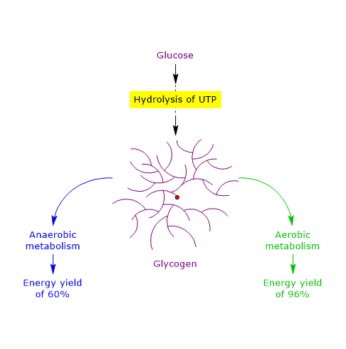
Under aerobic conditions, the overall energy retention efficiency of glycogen storage is 96%, demonstrating that glycogen is an exceptionally efficient molecular energy reserve, providing a 36% higher relative yield compared to anaerobic pathways.[51]
| Pathway/Condition | Initial cleavage step | Net ATP yield (per glucose unit) | ATP Saved in hexokinase step | Energy conservation efficiency (%) |
|---|---|---|---|---|
| Aerobic glycogen oxidation | Phosphorolytic (phosphorylase) | 31–33 ATP | 1 ATP | ≈ 96% |
| Anaerobic glycogenolysis/Glycolysis | Phosphorolytic (phosphorylase) | 3 ATP | 1 ATP | ≈ 60% |
| Free Glucose oxidation (comparison) | Hydrolytic/Hexokinase | 30–32 ATP (aerobic)/2 ATP (anaerobic) | 0 ATP | Lower overall efficiency |
References
- a b Daghlas S.A., Mohiuddin S.S. Biochemistry, Glycogen. [Updated 2023 May 1]. In: StatPearls [Internet]. Treasure Island (FL): StatPearls Publishing; 2025 Jan-. Available from: https://www.ncbi.nlm.nih.gov/books/NBK539802/
- a b c d e f Roach P.J., Depaoli-Roach A.A., Hurley T.D, Tagliabracci V.C. Glycogen and its metabolism: some new developments and old themes. Biochem J 2012;441:763-87. doi:10.1042/BJ20111416
- a b c d e f g Neoh G.K.S., Tan X., Chen S., Roura E., Dong X., Gilbert R.G. Glycogen metabolism and structure: a review. Carbohydr Polym 2024;346:122631. doi:10.1016/j.carbpol.2024.122631
- ^ Michal G., Schomburg D. Biochemical pathways. An atlas of biochemistry and molecular biology. 2nd Edition. John Wiley & Sons, Inc. 2012
- a b c d e f Prats C., Graham T.E., and Shearer J. The dynamic life of the glycogen granule. J Biol Chem 2018;293(19):7089-98. doi:10.1074/jbc.R117.802843
- a b c Adeva-Andany M.M., González-Lucán M., Donapetry-García C., Fernández-Fernández C., Ameneiros-Rodríguez E. Glycogen metabolism in humans. BBA Clin 2016;27;5:85-100. doi:10.1016/j.bbacli.2016.02.001
- a b Canibano-Fraile R., Harlaar L., Dos Santos C.A., et al. Lysosomal glycogen accumulation in Pompe disease results in disturbed cytoplasmic glycogen metabolism. J Inherit Metab Dis 2023;46(1):101-115. doi:10.1002/jimd.12560
- a b c d e f Stipanuk M.H., Caudill M.A. Biochemical, physiological, and molecular aspects of human nutrition. 4th Edition. St. Louis: Elsevier, 2018.
- a b c d e f g h i Nelson D.L., Cox M.M. Lehninger. Principles of biochemistry. 8th Edition. W.H. Freeman and Company, 2021.
- ^ Terlouw E.M.C., Picard B., Deiss V., Berri C., Hocquette J.F., Lebret B., Lefèvre F., Hamill R., Gagaoua M. Understanding the determination of meat quality using biochemical characteristics of the muscle: stress at slaughter and other missing keys. Foods 2021;10(1):84. doi:10.3390/foods10010084
- ^ Claude Bernard, the founder of modern medicine. Cells 2022;11(10):1702. doi:10.3390/cells11101702
- ^ The Nobel Foundation. Retrieved October 30, 2025. https://www.nobelprize.org/the-nobel-prize-organisation/the-nobel-foundation/
- a b c d Berg J.M., Tymoczko J.L., Gatto G.J., Stryer L. Biochemistry. 9th Edition. W.H. Freeman and Company, 2019.
- a b c d e f g Heilman D., Woski S., Voet D., Voet J.G., Pratt C.W. Fundamentals of biochemistry: life at the molecular level. 6th Edition. Wiley, 2023.
- a b c Garrett R.H., Grisham C.M. Biochemistry. 7th Edition. Brooks/Cole, Cengage Learning, 2023.
- ^ Alberts B., Johnson A., Lewis J., Morgan D., Raff M., Roberts K., Walter P. Molecular Biology of the Cell. 7th Edition. Garland Science, Taylor & Francis Group, 2022.
- ^ Yu Y., Delbianco M. Conformational studies of oligosaccharides. Chemistry 2020;26(44):9814-9825. doi:10.1002/chem.202001370
- a b Gunja-Smith Z., Marshall J.J., Mercier C., Smith E.E., Whelan W.J. A revision of the Meyer-Bernfeld model of glycogen and amylopectin. FEBS Lett 1970;12(2):101-104. doi:10.1016/0014-5793(70)80573-7
- ^ Whelan W.J. Enzymic explorations of the structures of starch and glycogen. Biochem J 1971;122(5):609-622. doi:10.1042/bj1220609
- ^ Meléndez-Hevia E., Waddell T.G., Shelton E.D. Optimization of molecular design in the evolution of metabolism: the glycogen molecule. Biochem J 1993;295(2):477-83. doi:10.1042/bj2950477
- ^ Shearer J., Graham T.E. Novel aspects of skeletal muscle glycogen and its regulation during rest and exercise. Exerc Sport Sci Rev 2004;32(3):120-6. doi:10.1097/00003677-200407000-00008
- ^ Marchand I., Chorneyko K., Tarnopolsky M., Hamilton S., Shearer J., Potvin J., Graham T.E. Quantification of subcellular glycogen in resting human muscle: granule size, number, and location. J Appl Physiol (1985) 2002;93(5):1598-607. doi:10.1152/japplphysiol.00585.2001
- ^ Meléndez R., Meléndez-Hevia E., Mas F., Mach J., Cascante M. Physical constraints in the synthesis of glycogen that influence its structural homogeneity: a two-dimensional approach. Biophys J 1998;75(1):106-14. doi:10.1016/S0006-3495(98)77498-3
- ^ Melendez R., Melendez-Hevia E., Cascante M. How did glycogen structure evolve to satisfy the requirement for rapid mobilization of glucose? A problem of physical constraints in structure building. J Mol Evol 1997;45(4):446-55. doi:10.1007/PL00006249
- ^ Fastman N.M., Liu Y., Ramanan V., Merritt H., et al. The structural mechanism of human glycogen synthesis by the GYS1-GYG1 complex. Cell Rep 2022;40(1):111041. doi:10.1016/j.celrep.2022.111041
- ^ Fontana J.D. The presence of phosphate in glycogen. FEBS Lett 1980;109(1):85-92. doi:10.1016/0014-5793(80)81317-2
- ^ Gentry M.S., Guinovart J.J., Minassian B.A., Roach P.J., Serratosa J.M. Lafora disease offers a unique window into neuronal glycogen metabolism. J Biol Chem 2018;293(19):7117-25. doi:10.1074/jbc.R117.803064
- ^ Turnbull J., Tiberia E., Pereira S., et al. Deficiency of a glycogen synthase-associated protein, Epm2aip1, causes decreased glycogen synthesis and hepatic insulin resistance. J Biol Chem 2013;288(48):34627-37. doi:10.1074/jbc.M113.483198
- ^ Roach P.J. Glycogen phosphorylation and Lafora disease. Mol Aspects Med 2015;46:78-84. doi:10.1016/j.mam.2015.08.003
- ^ Brewer M.K., Gentry M.S. Brain glycogen structure and its associated proteins: past, present and future. Adv Neurobiol 2019;23:17-81. doi:10.1007/978-3-030-27480-1_2
- ^ Sullivan M.A., Aroney S.T., Li S., Warren FJ, et al. Changes in glycogen structure over feeding cycle sheds new light on blood-glucose control. Biomacromolecules 2014;15(2):660-5. doi:10.1021/bm401714v
- ^ Baqué S., Guinovart J.J., Ferrer J.C. Glycogenin, the primer of glycogen synthesis, binds to actin. FEBS Lett 1997;417(3):355-9. doi:10.1016/s0014-5793(97)01299-4
- ^ Katz J., Tayek J.A. Gluconeogenesis and the Cori cycle in 12-, 20-, and 40-h-fasted humans. Am J Physiol 1998;275(3):E537-42. doi:10.1152/ajpendo.1998.275.3.E537
- ^ Felig P., Pozefsk T., Marlis E., Cahill G.F. Alanine: key role in gluconeogenesis. Science 1970;167(3920):1003-1004. doi:10.1126/science.167.3920.1003
- ^ Wilson J.E. Isozymes of mammalian hexokinase: structure, subcellular localization and metabolic function. J Exp Biol 2003;206(Pt 12):2049-57. doi:10.1242/jeb.00241
- ^ Horecker B.L. The pentose phosphate pathway. J Biol Chem 2002;277(50):47965-47971. doi:10.1074/jbc.X200007200
- ^ Smythe C., Cohen P. The discovery of glycogenin and the priming mechanism for glycogen biogenesis. Eur J Biochem 1991;200(3):625-31. doi:10.1111/j.1432-1033.1991.tb16225.x
- ^ Zois C.E., Harris A.L. Glycogen metabolism has a key role in the cancer microenvironment and provides new targets for cancer therapy. J Mol Med (Berl) 2016;94(2):137-54. doi:10.1007/s00109-015-1377-9
- ^ Mandl J. Glycogen-endoplasmic reticulum connection in the liver. Int J Mol Sci 2023;24(2):1074. doi:10.3390/ijms24021074
- a b Rajas F., Gautier-Stein A., Mithieux G. Glucose 6-phosphate, a central hub for liver carbohydrate metabolism. Metabolites 2019;9(12):282. doi:10.3390/metabo9120282
- ^ Zirin J., Nieuwenhuis J., Perrimon N. Role of autophagy in glycogen breakdown and its relevance to chloroquine myopathy. PLoS Biol 2013;11(11):e1001708. doi:10.1371/journal.pbio.1001708
- ^ Koutsifeli P., Varma U., Daniels L.J., et al. Glycogen-autophagy: molecular machinery and cellular mechanisms of glycophagy. J Biol Chem 2022;298(7):102093. doi:10.1016/j.jbc.2022.102093
- ^ Okuyama M., Saburi W., Mori H., Kimura A. α-Glucosidases and α-1,4-glucan lyases: structures, functions, and physiological actions. Cell Mol Life Sci 2016;73(14):2727-51. doi:10.1007/s00018-016-2247-5
- ^ Rahman M.S., Hossain K.S., Das S., et al. Role of insulin in health and disease: an update. Int J Mol Sci 2021;22(12):6403. doi:10.3390/ijms22126403
- ^ Knuiman P., Hopman M.T., Mensink M. Glycogen availability and skeletal muscle adaptations with endurance and resistance exercise. Nutr Metab (Lond) 2015;12:59. doi:10.1186/s12986-015-0055-9
- ^ Beelen M., Burke L.M., Gibala M.J., van Loon J.C. Nutritional strategies to promote postexercise recovery. Int J Sport Nutr Exerc Metab 2010;20(6):515-32. doi:10.1123/ijsnem.20.6.515
- ^ Farrell P.M., Bourbon J.R. Fetal lung surfactant lipid synthesis from glycogen during organ culture. Biochim Biophys Acta 1986;878(2):159-67. doi:10.1016/0005-2760(86)90142-6
- ^ Yildiz Atar H., Baatz J.E., Ryan R.M. Molecular mechanisms of maternal diabetes effects on fetal and neonatal surfactant. Children (Basel) 2021;8(4):281. doi:10.3390/children8040281
- ^ Rosenthal M.D., Glew R.H. Medical biochemistry − Human metabolism in health and disease. John Wiley & Sons, Inc., 2009.
- ^ Duran J., Gruart A., López-Ramos J.C., Delgado-García J.M., Guinovart J.J. Glycogen in astrocytes and neurons: physiological and pathological aspects. Adv Neurobiol 2019;23:311-329. doi:10.1007/978-3-030-27480-1_10
- a b c d Snider M.D., Croniger C.M. Devlin′s textbook of biochemistry with clinical correlations. 8th Edition. John Wiley & Sons, Inc. 2024.
Domande Frequenti
What is the difference between liver and muscle glycogen reserves?
Liver glycogen (≈ 100 g) releases free glucose into blood via glucose 6-phosphatase for glycemic homeostasis. Muscle glycogen (250-300 g), since muscle lacks phosphatase, serves exclusively as a private energy substrate for local ATP production.
Why does glycogen breakdown save energy compared to free glucose?
Glycogen phosphorylase performs phosphorolytic cleavage producing glucose 1-phosphate, converted to glucose 6-phosphate without energy cost. This bypasses the hexokinase ATP-consuming step, increasing net glycolytic ATP yield by 1 molecule.
What is the essential role of glycogenin in glycogen synthesis?
Glycogenin is a self-glicosylating enzyme that acts as an obligate primer for glycogenesis. It synthesizes an initial short chain of 8 glucose residues with α-(1→4) glycosidic bonds, creating the foundation required by glycogen synthase.