Category Archives: Phytochemicals

Phytochemicals are organic compounds, generally of low molecular weight, produced by plants. Based on the structural characteristics they can be divided into three groups: polyphenols, carotenoids and glucosinolates.
They are produced by primary and secondary plant metabolism, and in plant they play many roles, some of which are listed below.

  • They are important during growth.
  • They play a role in defense against predators, such as herbivores, competitors, and pathogenic fungi, bacteria and viruses.
  • They protect from environmental agents such as UV rays.
  • Considering edible vegetables, they contribute to organoleptic characteristics such as color, flavor and aroma.

Phytochemicals are not essential nutrients for humans.
They are present in vegetables, fruits, cereals, legumes, and derived products, such as wine, tea, chocolate or extra virgin olive oil. It is believed that the beneficial effects on health ascribed by scientific literature and tradition to the consumption of these foods are also due to their presence.
Phytochemicals protect against many diseases. Although some are toxic to humans, namely, they are phytotoxins, others act as antinutrients interfering with the absorption of some nutrients, interventional and observational studies have shown that many of them are believed to be effective in preventing chronic diseases such as cardiovascular disease, many type of cancers or neurodegenerative diseases.
How do they work at the molecular level?
After binding to membrane and/or intracellular receptors, they activate specific signaling pathways. This modify the activity of some enzymes and/or induce the phosphorylation of transcription factors, then influencing gene expression. And, depending on the target cell, specific cellular processes will be activated or inhibited.
Should phytochemicals be considered drugs? The low bioavailability and their metabolism cause these compounds and their metabolites to be present in the circulation in very low concentrations, far from those required for pharmacological action. To this it should be added that they have cytotoxic effects in pharmacological doses.

Flavonoid biosynthesis in plants: genes and enzymes

Flavonoid biosynthesis, probably the best characterized pathway of plant secondary metabolism, is part of the phenylpropanoid pathway that, in addition to flavonoids, leads to the formation of a wide range of phenolic compounds, such as hydroxycinnamic acids, stilbenes, lignans and lignins.
Flavonoid biosynthesis is linked to primary metabolism through both mitochondria- and plastid-derived molecules. Since it seems that most of the involved enzymes characterized to date operate in protein complexes located in the cell cytosol, these molecules must be exported to the cytoplasm to be used.
The end products are transported to different intracellular or extracellular locations, with flavonoids involved in pigmentation usually transported into the vacuoles.
The biosynthesis of this group of polyphenols requires one p-coumaroyl-CoA and three malonyl-CoA molecules as initial substrates.
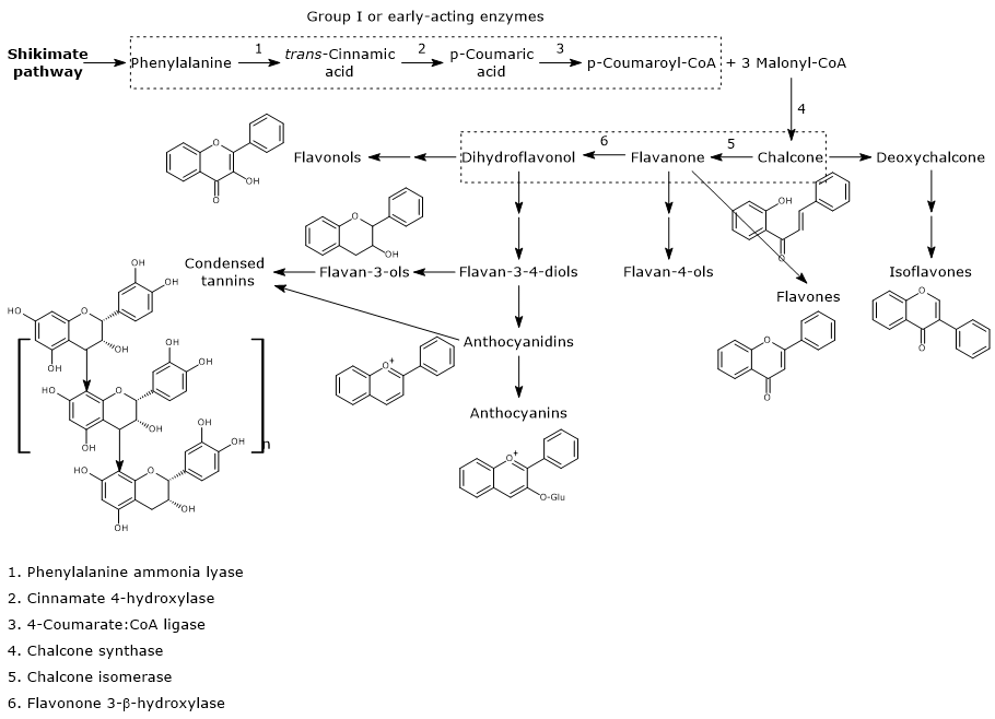
Flavonoid biosynthesis pathway
Flavonoid Biosynthesis

Contents

Biosynthesis of p-coumaroyl-CoA

p-Coumaroyl-CoA is the pivotal branch-point metabolite in the phenylpropanoid pathway, being the precursor of a wide variety of phenolic compounds, both flavonoid and non-flavonoid polyphenols.
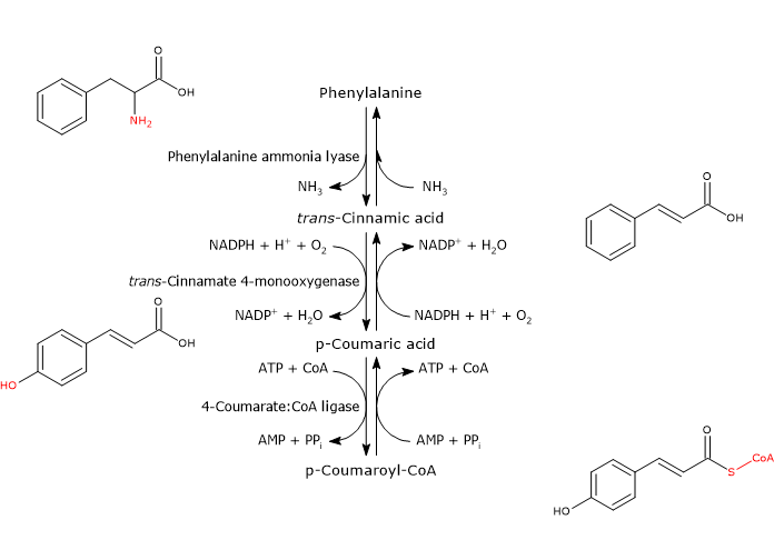
It is produced from phenylalanine via three reactions catalyzed by cytosolic enzymes collectively called group I or early-acting enzymes, in order of action:

  • phenylalanine ammonia lyase (EC 4.3.1.24);
  • trans-cinnamate 4-monooxygenase (EC:1.14.14.91);
  • 4-coumarate-CoA ligase (EC 6.2.1.12).
Biosynthesis of p-coumaroyl-CoA from phenylalanine
Biosynthesis of p-coumaroyl-CoA

They seems to be associated in a multienzyme complex anchored to the endoplasmic reticulum membrane. The anchoring is probably ensured by cinnamate 4-hydroxylase that inserts its N-terminal domain into the membrane of the endoplasmic reticulum itself. These complexes, referred to as “metabolons”, allow the product of a reaction to be channeled directly as substrate to the active site of the enzyme that catalyzes the consecutive reaction in the metabolic pathway.
With the exception of cinnamate 4-hydroxylase, the enzymes which act downstream of phenylalanine ammonia lyase are encoded by small gene families in all species analyzed so far.
The different isoenzymes show distinct temporal, tissue, and elicitor-induced patterns of expression. It seems, in fact, that each member of each family can be used mainly for the synthesis of a specific compound, thus acting as a control point for carbon flux among the metabolic pathways leading to lignan, lignin, and flavonoid biosynthesis.

Note: Phenylalanine is a product of the shikimic acid pathway, which converts simple precursors derived from the metabolism of carbohydrates, phosphoenolpyruvate and erythrose-4-phosphate, intermediates of glycolysis and the pentose phosphate pathway, respectively, into the aromatic amino acids phenylalanine, tyrosine and tryptophan. Unlike plants and microorganisms, animals do not possess the shikimic acid pathway, and are not able to synthesize the three above-mentioned amino acids, which are therefore essential nutrients.

Phenylalanine ammonia lyase (PAL)

It is one of the most studied and best characterized enzymes of plant secondary metabolism. It requires no cofactors and catalyzes the reaction that links primary and secondary metabolism: the reversible deamination of phenylalanine to trans-cinnamic acid, with the release of nitrogen as ammonia and introduction of a trans double bond between carbon atoms 7 and 8 of the side chain.

Phenylalanine ⇄ trans-Cinnamic Acid + NH3

Therefore, it directs the flow of carbon from the shikimic acid pathway to the different branches of the phenylpropanoid pathway. The released ammonia is probably fixed in the reaction catalyzed by glutamine synthetase.
The enzyme from monocots is also able to act as tyrosine ammonia lyase (EC 4.3.1.25), converting tyrosine to p-coumaric acid directly, (therefore without the 4-hydroxylation step), but with a lower efficiency.
In all plant species investigated, several copies of phenylalanine ammonia lyase gene are found, copies that probably respond differentially to internal and external stimuli. Indeed, gene transcription, and then enzyme activity, are under the control of both internal developmental and external environmental stimuli. Here are some examples that require increased enzyme activity.

  • The flowering.
  • The production of lignin to strengthen the secondary wall of xylem cells.
  • The production of flower pigments that attract pollinators.
  • Pathogen infections, that require the production of phenylpropanoid phytoalexins, or exposure to UV rays.

trans-Cinnamate 4-monooxygenase

It belongs to the cytochrome P450 superfamily (EC 1.14.-.-), is a microsomal monooxygenase containing a heme cofactor, and dependent on both O2 and NADPH. It catalyzes the formation of p-coumaric acid through the introduction of a hydroxyl group in 4-position of trans-cinnamic acid (this hydroxyl group is present in most flavonoids).

trans-Cinnamic Acid + NADPH + H+ + O2 ⇄ p-Coumaric Acid + NADP+ + H2O

This reaction is also part of the biosynthesis of hydroxycinnamic acids.
Increases in transcription rates and enzyme activity are observed in correlation with the synthesis of phytoalexins (in response to fungal infections), lignification as well as wounding.

4-Coumarate:CoA ligase (4CL)

With Mg2+ as a cofactor, it catalyzes the ATP-dependent activation of the carboxyl group of p-coumaric acid and other hydroxycinnamic acids, metabolically rather inert molecules, through the formation of the corresponding CoA-thioester.

p-Coumaric Acid + ATP + CoA ⇄ p-Coumaroyl-CoA + AMP + PPi

Generally, p-coumaric acid and caffeic acid are the preferred substrates, followed by ferulic acid and 5-hydroxyferulic acid, low activity against trans-cinnamic acid and none against sinapic acid. These CoA-thioesters are able to enter various reactions such as:

  • reduction to alcohol (monolignols) or aldehydes;
  • stilbene and flavonoid biosynthesis;
  • transfer to acceptor molecules.

It should finally be pointed out that the activation of the carboxyl group can also be obtained through an UDP-glucose-dependent transfer to glucose.

Biosynthesis of malonyl-CoA

Malonyl-CoA does not derived from the phenylpropanoid pathway, but from the reaction catalyzed by acetyl-CoA carboxylase (EC 6.4.1.2, the cytosolic form, see below). The enzyme, with biotin and Mg2+ as cofactors, catalyzes the ATP-dependent carboxylation of acetyl-CoA, using bicarbonate as a source of carbon dioxide (CO2).

Acetyl-CoA + HCO3 + ATP → Malonyl-CoA + ADP + Pi

It is found both in the plastids, where it participates in the synthesis of fatty acids, and the cytoplasm, and is the latter that catalyzes the formation of malonyl-CoA that is used in the biosynthesis of flavonoids and other compounds. Increases in the transcription rate of the gene and enzyme activity are induced in response to stimuli that increase the biosynthesis of these polyphenols, such as exposure to pathogenic fungi or UV-rays.
In turn, acetyl-CoA is produced in plastids, mitochondria, peroxisomes and cytosol through different metabolic pathways. The molecules used in the biosynthesis of malonyl-CoA, and therefore of the flavonoids, are the cytosolic ones, produced in the reaction catalyzed by ATP-citrate lyase (EC 2.3.3.8) that cleaves citrate, in the presence of CoA and ATP, to form oxaloacetate and acetyl-CoA, plus ADP and inorganic phosphate.

First steps in flavonoid biosynthesis

The first step in flavonoid biosynthesis is catalyzed by chalcone synthase (EC 2.3.1.74), an enzyme anchored to the endoplasmic reticulum and with no known cofactors.
From one p-coumaroyl-CoA and three malonyl-CoA, it catalyzes sequential condensation and decarboxylation reactions in the course of which a polyketide intermediate is formed. The polyketide undergoes cyclizations and aromatizations leading to the formation of the A ring. The product of the reactions is naringenin chalcone (2′,4,4′,6′-tetrahydroxychalcone), a 6′-hydroxychalcone and the first flavonoid to be synthesized by plants.

p-Coumaroyl-CoA + 3 Malonyl-CoA → Naringenin Chalcone + 4 CoA + 3 CO2

The reaction, cytosolic, is irreversible due to the release of three CO2 and 4 CoA.
B ring and the three-carbon bridge of the molecule originate from p-coumaroyl-CoA (and therefore from phenylalanine), the A ring from the three malonyl-CoA units.
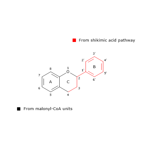
Flavonoid biosynthesis and the origin of the flavonoid skeleton
The Origin of the Flavonoid Skeleton

Also 6’-deoxychalcone can be produced; its synthesis is thought to involve an additional reduction step catalyzed by polyketide reductase (EC. 1.1.1.-).
Chalcone synthase from some plant species, such as barley (Hordeum vulgare), accepts as substrates also caffeoil-CoA, feruloil-CoA and cinnamoyl-CoA.
It is the most abundant enzyme of the phenylpropanoid pathway, probably because it has a low catalytic activity, and, in fact, is considered to be the rate-limiting enzyme in flavonoid biosynthesis.
As for phenylalanine ammonia lyase, chalcone synthase gene expression is under the control of both internal and external factors. In some plants, one or two isoenzymes are found, while in others up to 9.
Chalcone synthase belongs to polyketide synthase group, present in bacteria, fungi and plants. These enzymes are able to catalyze the production of polyketide chains through sequential condensations of acetate units provided by malonyl-CoA units. They also includes stilbene synthase (EC 2.3.1.146), which catalyzes the formation of resveratrol, a non flavonoid polyphenol compound that has attracted much interest because of its potential health benefits.
Generally, chalcones do not accumulate in plants because naringenin chalcone is converted to (2S)-naringenin, a flavanone, in the reaction catalyzed by chalcone isomerase (EC 5.5.1.6).
The enzyme, the first of the flavonoid biosynthesis to be discovered, catalyzes a stereospecific isomerization and closes the C ring. Two types of chalcone isomerases are known, called type I and II. Type I enzymes use only 6′-hydroxychalcone substrates, like naringenin chalcone, while type II, prevalent in legumes, use both 6′-hydroxy- and 6′-deoxychalcone substrates.
It should be noted that with 6′-hydroxychalcones, isomerization can also occur nonenzymically to form a racemic mixture, both in vitro and in vivo, enough to allow a moderate synthesis of anthocyanins. On the contrary, under physiological conditions 6′-deoxychalcones are stable, and so the activity of type II chalcone isomerases is required to form flavanones.
The enzyme increases the rate of the reaction of 107 fold compared to the spontaneous reaction, but with a higher kinetics for the 6′-hydroxychalcones than 6′-deoxychalcones. Finally, it produces (2S)-flavanones, which are the biosynthetically required enantiomers.
As other enzymes in flavonoid biosynthesis, also chalcone isomerase gene expression is subject to strict control. And, as phenylalanine ammonia lyase and chalcone synthase, it is induced by elicitors.
In the reaction catalysed by flavanone-3β-hydroxylase (EC 1.14.11.9), (2S)-flavanones undergo a stereospecific isomerization that converts them into the respective (2R,3R)-dihydroflavonols. In particular, naringenin is converted into dihydrokaempferol.
The enzyme is a cytosolic non-heme-dependent dioxygenase, dependent on Fe2+ and 2-oxoglutarate, and therefore belonging to the family of 2-oxoglutarate-dependent dioxygenase (which distinguishes them from the other hydroxylases of the flavonoid biosynthetic pathway which are cytochrome P450 enzymes).
Naringenin chalcone, (2S)-naringenin, and dihydrokaempferol are central intermediates in flavonoid biosynthesis, since they act as branch-point compounds from which the synthesis of distinct flavonoid subclasses can occur. For example, directly or indirectly:

Not all of these side metabolic pathways are present in every plant species, or are active within each tissue type of a given plant. Like enzymes previously seen, the activity of those involved in these “side-routes” is subject to strict control, resulting in a tissue-specific profile of flavonoid compounds. For example, grape seeds, flesh and skin have distinct anthocyanin, catechin, flavonol and condensed tannin profiles, whose synthesis and accumulation are strictly and temporally coordinated during the ripening process.

References

  1. Andersen Ø.M., Markham K.R. Flavonoids: chemistry, biochemistry, and applications. CRC Press Taylor & Francis Group, 2006
  2. de la Rosa L.A., Alvarez-Parrilla E., Gonzàlez-Aguilar G.A. Fruit and vegetable phytochemicals: chemistry, nutritional value, and stability. 1th Edition. Wiley J. & Sons, Inc., Publication, 2010
  3. Heldt H-W. Plant biochemistry – 3th Edition. Elsevier Academic Press, 2005
  4. Vogt T. Phenylpropanoid biosynthesis. Mol Plant 2010;3(1):2-20. doi:10.1093/mp/ssp106
  5. Wink M. Biochemistry of plant secondary metabolism – 2nd Edition. Annual plant reviews (v. 40), Wiley J. & Sons, Inc., Publication, 2010
Lignans: structure, metabolism, benefits, and sources

Lignans are a subgroup of non-flavonoid polyphenols.
They are widely distributed in the plant kingdom, being present in more than 55 plant families, where they act as antioxidants and defense molecules against pathogenic fungi and bacteria.
In humans, epidemiological and physiological studies have shown that they can exert positive effects in the prevention of lifestyle-related diseases, such as type II diabetes and cancer. For example, an increased dietary intake of these polyphenols correlates with a reduction in the occurrence of certain types of estrogen-related tumors, such as breast cancer in postmenopausal women.
In addition, some lignans have also aroused pharmacological interest. Examples are:

  • podophyllotoxin, obtained from plants of the genus Podophyllum (Berberidaceae family); it is a mitotic toxin whose derivatives have been used as chemotherapeutic agents;
  • arctigenin and tracheologin, obtained from tropical climbing plants; they have antiviral properties and have been tested in the search for a drug to treat AIDS .

Contents

Chemical structure

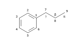
Their basic chemical structure consists of two phenylpropane units linked by a C-C bond between the central atoms of the respective side chains (position 8 or β), also called β-β’ bond. 3-3′, 8-O-4′, or 8-3′ bonds are observed less frequently; in these cases the dimers are called neolignans. Hence, their chemical structure is referred to as (C6-C3)2, and they are included in the phenylpropanoid group, as well as their precursors: the hydroxycinnamic acids.

Skeletal formula of phenylpropanoid unit of lignans
Phenylpropanoid unit

Based on their carbon skeleton, cyclization pattern, and the way in which oxygen is incorporated in the molecule skeleton, they can be divided into 8 subgroups: furans, furofurans, dibenzylbutanes, dibenzylbutyrolactones, dibenzocyclooctadienes, dibenzylbutyrolactols, aryltetralins and arylnaphthalenes. Furthermore, there is considerable variability regarding the oxidation level of both the propyl side chains and the aromatic rings.
They are not present in the free form in nature, but linked to other molecules, mainly as glycosylated derivatives.
Among the most common lignans, secoisolariciresinol (the most abundant one), lariciresinol, pinoresinol, matairesinol and 7-hydroxymatairesinol are found.

Note: They occur not only as dimers but also as more complex oligomers, such as dilignans and sesquilignans.

Biosynthesis

In this section, we will examine the biosynthesis of some of the most common lignans.
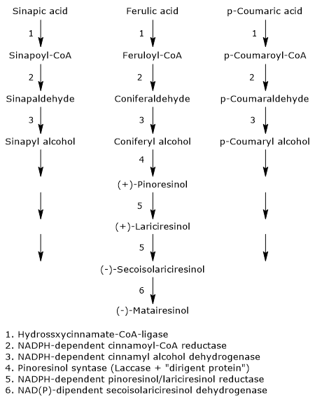
The pathway starts from 3 of the 4 most common dietary hydroxycinnamic acids: p-coumaric acid, sinapic acid, and ferulic acid (caffeic acid is not a precursor of this subgroup of polyphenols). Therefore, they arise from the shikimic acid pathway, via phenylalanine.

Synthesis pathways for lignans
Lignan Biosynthesis

The first three reactions reduce the carboxylic group of the hydroxycinnamates to alcohol group, with formation of the corresponding alcohols, called monolignols, that is, p-coumaric alcohol, sinapyl alcohol and coniferyl alcohol. These molecules also enter the pathway of lignin biosynthesis.

  • The first step, which leads to the activation of the hydroxycinnamic acids, is catalysed by hydroxycinnamate:CoA ligases, commonly called p-coumarate:CoA ligases (EC 6.2.1.12), with formation of the corresponding hydroxycinnamate-CoAs, namely, feruloil-CoA, p- coumaroyl-CoA and sinapil-CoA.
  • In the second step, a NADPH-dependent cinnamoyl-CoA: oxidoreductase, also called cinnamoyl-CoA reductase (EC1.2.1.44) catalyzes the formation of the corresponding aldehydes, and the release of coenzyme A.
  • In the last step, a NADPH-dependent cinnamyl alcohol dehydrogenase, also called monolignol dehydrogenase (EC 1.1.1.195), catalyzes the reduction of the aldehyde group to an alcohol group, with the formation of the aforementioned monolignols.

The next step, the dimerization of monolignols, involves the intervention of stereoselective mechanisms, or, more precisely, enantioselective mechanisms.In fact, most of the plant lignans exists as (+)- or (-)-enantiomers, that is, isomers with property of chirality, whose relative amounts can vary from species to species, but also in different organs on the same plant, depending on the type of reactions involved.
The dimerization can occur through enzymatic reactions involving laccases (EC 1.10.3.2). These enzymes catalyze the formation of radicals that, dimerizing, form a racemic mixture. However, this does not explain how the racemic mixtures found in plants are formed. The most accepted mechanism to explain the stereospecific synthesis involves the action of the laccase and of a protein able to direct the synthesis toward one or the other of the two enantiomeric forms: the dirigent protein. The reaction scheme might be: the enzyme catalyzes the synthesis of phenylpropanoid radicals that are orientated in such a way to obtain the desired stereospecific coupling by the dirigent protein.

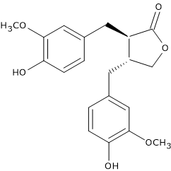
Skeletal formula of the lignan (-)-matairesinol
(-)-Matairesinol

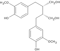
For example, pinoresinol synthase, consisting of laccase and dirigent protein, catalyzes the stereospecific synthesis of (+)-pinoresinol from two units of coniferyl alcohol. (+)-Pinoresinol, in two consecutive stereospecific reactions catalyzed by NADPH-dependent pinoresinol/lariciresinol reductase (EC 1.23.1.2), is first reduced to (+)-lariciresinol, and then to (-)-secoisolariciresinol. (-)-Secoisolariciresinol, in the reaction catalyzed by NAD(P)-dependent secoisolariciresinol dehydrogenase (EC 1.1.1.331) is oxidized to (-)-matairesinol.

Metabolism by human gut microbiota

Their importance to human health is due largely to their metabolism by gut microbiota, which is part of the larger human microbiota, and that carries out deglycosylations, para-dehydroxylations, and meta-demethylations without enantiomeric inversion. Indeed, this metabolization leads to the formation molecules with a modest estrogen-like activity (phytoestrogens), a situation similar to that observed with some isoflavones, such as those of soybean, some coumarins, and some stilbenes. These active metabolites are the so-called “mammalian lignans or enterolignans”, such as the aglycones of enterodiol and enterolactone, formed from secoisolariciresinol and matairesinol, respectively.
Studies conducted on animals fed diets rich in lignans have shown their presence as intact molecules, in low concentrations, in serum, suggesting that they may be absorbed as such from the intestine. These molecules exhibit estrogen-independent actions, both in vivo and in vitro, such as inhibition of angiogenesis, reduction of diabetes, and suppression of tumor growth.
Note: The term “phytoestrogen” refers to molecules with estrogenic or antiandrogenic activity, at least in vitro.

Once absorbed, they enter the enterohepatic circulation, and, in the liver, may undergo phase II reactions and be sulfated or glucuronidated, and finally excreted in the urine.

Food sources

The richest dietary source is flaxseed (linseed), that contains mainly secoisolariciresinol, but also lariciresinol, pinoresinol and matairesinol in good quantity (for a total amount of more than 3.7 mg/100 g dry weight). They are also found in sesame seeds.

Skeletal formula of the lignan (-)-secoisolariciresinol
(-)-Secoisolariciresinol

Another important source is whole grains.
They are also present in other foods, but in concentrations from one hundred to one thousand times lower than those of flaxseed. Examples are:

  • beverages, generally more abundant in red wine, followed in descending order by black tea, soy milk and coffee;
  • fruits, such as apricots, pears, peaches, strawberries;
  • among vegetables, Brassicaceae, garlic, asparagus and carrots;
  • lentils and beans.

Their presence in grains and, to a lesser extent in red wine and fruit, makes them, at least in individuals who follow a mediterranean diet, the main source of phytoestrogens.

References

  1. Andersen Ø.M., Markham K.R. Flavonoids: chemistry, biochemistry, and applications. CRC Press Taylor & Francis Group, 2006
  2. de la Rosa L.A., Alvarez-Parrilla E., Gonzàlez-Aguilar G.A. Fruit and vegetable phytochemicals: chemistry, nutritional value, and stability. 1th Edition. Wiley J. & Sons, Inc., Publication, 2010
  3. Heldt H-W. Plant biochemistry – 3th Edition. Elsevier Academic Press, 2005
  4. Manach C., Scalbert A., Morand C., Rémésy C., and Jime´nez L. Polyphenols: food sources and bioavailability. Am J Clin Nutr 2004;79(5):727-747. doi:10.1093/ajcn/79.5.727
  5. Satake H, Koyama T., Bahabadi S.E., Matsumoto E., Ono E. and Murata J. Essences in metabolic engineering of lignan biosynthesis. Metabolites 2015;5:270-290. doi:10.3390/metabo5020270
  6. Tsao R. Chemistry and biochemistry of dietary polyphenols. Nutrients 2010;2:1231-1246. doi:10.3390/nu2121231
  7. van Duynhoven J., Vaughan E.E., Jacobs D.M., Kemperman R.A., van Velzen E.J.J, Gross G., Roger L.C., Possemiers S., Smilde A.K., Doré J., Westerhuis J.A.,and Van de Wiele T. Metabolic fate of polyphenols in the human superorganism. PNAS 2011;108(suppl. 1):4531-4538. doi:10.1073/pnas.1000098107
  8. Wink M. Biochemistry of plant secondary metabolism – 2nd Edition. Annual plant reviews (v. 40), Wiley J. & Sons, Inc., Publication, 2010
Hydroxycinnamic acids: structure, synthesis, health benefits, foods

Hydroxycinnamic acids or hydroxycinnamates are phenolic compounds belonging to non-flavonoid polyphenols.
They are present in all parts of fruits and vegetables although the highest concentrations are found in the outer part of ripe fruits, concentrations that decrease during ripening, while the total amount increases as the size of the fruits increases.
Their dietary intake has been associated with the prevention of:

  • cardiovascular diseases;
  • cancer;
  • type-2 diabetes.

These effects do seem to be due not only to their high antioxidant activity, that depends upon the hydroxylation pattern of the aromatic ring, but also to other mechanisms of action such as, e.g., the reduction of intestinal absorption of glucose or the modulation of secretion of some gut hormones.

Contents

Chemical structure

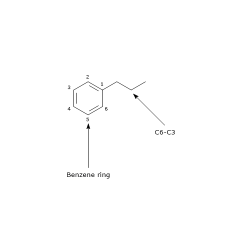
Their basic structure is a benzene ring to which a three carbon chain is attached, structure that is referred to as C6-C3. Therefore they can be included in the phenylpropanoid group.

Basic skeleton structure of hydroxycinnamic acids, phenolic compounds belonging to non-flavonoid polyphenols
Basic Skeleton of Hydroxycinnamates

The main dietary hydroxycinnamates are:

  • caffeic acid or 3,4-dihydroxycinnamic acid;
  • ferulic acid or 4-hydroxy-3-methoxycinnamic acid;
  • sinapic acid or 4-hydroxy-3,5-dimethoxycinnamic acid;
  • p-coumaric acid or 4-coumaric acid or 4-hydroxycinnamic acid.

In nature, they are associated with other molecules to form, e.g., glycosylated derivatives or esters of tartaric acid, quinic acid, or shikimic acid. In addition, several hundreds of anthocyanins acylated with the aforementioned hydroxycinnamates have been identified (in descending order with p-coumaric acid, more than 150, caffeic acid, about 100, ferulic acid, about 60, and sinapic acid, about 25). They are rarely present in the free form, except in processed foods that have undergone fermentation, sterilization or freezing. For example, an overlong storage of blood orange fruits causes a massive hydrolysis of hydroxycinnamic derivatives to free acids, and this in turn could lead to the formation of malodorous compounds such as vinyl phenols, indicators of too advanced senescence of the fruit.

Biosynthesis

Hydroxycinnamate biosynthesis consists of a series of enzymatic reactions subsequent to that catalyzed by phenylalanine ammonia lyase (PAL).

Phenylalanine ⇄ trans-Cinnamic acid + NH3

This enzyme catalyzes the deamination of phenylalanine to yield trans-cinnamic acid, so linking the aromatic amino acid to the hydroxycinnamic acids and their activated forms.

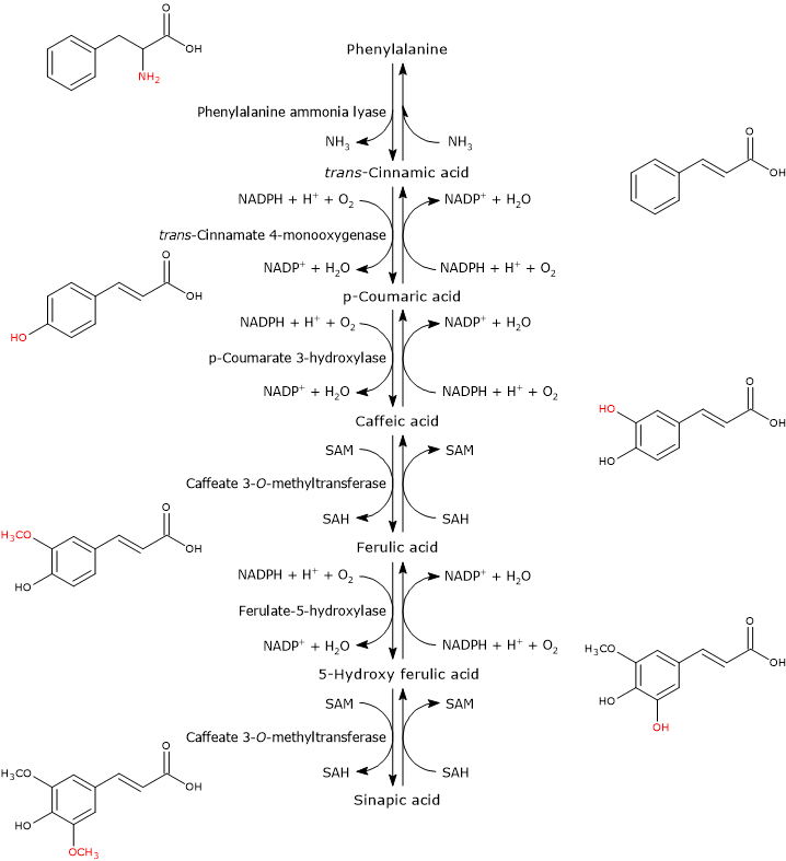
Synthesis of hydroxycinnamic acids from phenylalanine
Biosynthesis of Hydroxycinnamates

In the first step, a hydroxyl group is attached at the 4-position of the aromatic ring of trans-cinnamic acid to form p-coumaric acid. The reaction catalysed by trans-cinnamate 4-monooxygenase (EC:1.14.14.91).

trans-Cinnamic acid + NADPH + H+ + O2 ⇄ p-Coumaric acid + NADP+ + H2O

The addition of a second hydroxyl group at the 3-position of the ring of p-coumaric acid leads to the formation of caffeic acid. The reaction catalysed by p-coumarate 3-hydroxylase (EC 1.14.13.-).

p-Coumaric acid + NADPH + H+ + O2 ⇄ Caffeic acid + NADP+ + H2O

The O-methylation of the hydroxyl group at the 3-position yields ferulic acid. The reaction catalyzed by caffeate 3-O-methyltransferase (EC:2.1.1.68).

Caffeic acid + SAM ⇄ Ferulic acid + SAH

Ferulic acid is converted into sinapic acid through two reactions: a hydroxylation at the 5-position to form 5-hydroxy ferulic acid, in a reaction catalyzed by ferulate 5-hydroxylase (EC:1.14.-.-), and the subsequent O-methylation of the same hydroxyl group in a reaction catalyzed by caffeate 3-O-methyltransferase.

Ferulic acid + NADPH + H+ + O2 ⇄ 5-Hydroxy ferulic acid + NADP+ + H2O

5-Hydroxy ferulic acid + SA from M ⇄ Sinapic acid + SAH

Hydroxycinnamic acids are not present in high quantities since they are rapidly converted to glucose esters or coenzyme A (CoA) esters, in reactions catalyzed by O-glucosyltransferases and hydroxycinnamate:CoA ligases, respectively. These activated intermediates are branch points, being able to participate in a wide range of reactions such as condensation with malonyl-CoA to form flavonoids, or the NADPH-dependent reduction to form lignans, which are precursors of lignin.

Food sources

Kiwis, blueberries, plums, cherries, apples, pears, chicory, artichokes, carrots, lettuce, eggplant, wheat and coffee are among the richest sources.

Caffeic acid

It is generally, both in the free form and bound to other molecules, the most abundant hydroxycinnamic acid in vegetables and most of the fruits, where it represents between 75 and 100 percent of the hydroxycinnamates.
The richest sources are coffee (drink), carrots, lettuce, potatoes, even sweet ones, and berries such as blueberries, cranberries and blackberries.
Smaller quantities are present in grapes and grape-derived products, orange juice, apples, plums, peaches, and tomatoes.
Caffeic acid and quinic acid bind to form chlorogenic acid, present in many fruit and in high concentration in coffee.

Ferulic acid

It is the most abundant hydroxycinnamic acid in cereals, which are also its main dietary source.
In wheat grain, its content is between 0.8 and 2 g/kg dry weight, which represents up to 90 percent of the total polyphenols. It is found chiefly, up to 98 percent of the total content, in the aleurone layer and pericarp, namely, the outer parts of the grain, and therefore its content in wheat flours depends upon the degree of refining, while the main source is obviously the bran. The molecule is present mainly in the trans form, and esterified with arabinoxylans and hemicelluloses. And in fact, in wheat bran the soluble free form represents only about 10 percent of its total amount. Dimers were also found, which form bridge structures between chains of hemicellulose.
In fruits and vegetables, ferulic acid is much less common than caffeic acid. The main sources are asparagus, eggplant and broccoli; lower quantities are found in blackberries, blueberries, cranberries, apples, carrots, potatoes, beets, coffee and orange juice.

Sinapic acid

The highest amounts are found in citrus peel and seeds (in orange juice, the amount is much lower); appreciable quantities in Chinese cabbage and in some varieties of cranberries.

p-Coumaric acid

High amounts are present in eggplant, the richest source, broccoli and asparagus; other sources are sweet cherries, plums, blueberries, cranberries, citrus peel and seeds, and orange juice.

References

  1. Andersen Ø.M., Markham K.R. Flavonoids: chemistry, biochemistry, and applications. CRC Press Taylor & Francis Group, 2006
  2. de la Rosa L.A., Alvarez-Parrilla E., Gonzàlez-Aguilar G.A. Fruit and vegetable phytochemicals: chemistry, nutritional value, and stability. 1th Edition. Wiley J. & Sons, Inc., Publication, 2010
  3. Manach C., Scalbert A., Morand C., Rémésy C., and Jime´nez L. Polyphenols: food sources and bioavailability. Am J Clin Nutr 2004;79(5):727-747. doi:10.1093/ajcn/79.5.727
  4. Preedy V.R. Coffee in health and disease prevention. Academic Press, 2014
  5. Zhao Z., Moghadasian M.H. Bioavailability of hydroxycinnamates: a brief review of in vivo and in vitro studies. Phytochem Rev 2010;9(1):133-145. doi:10.1007/s11101-009-9145-5

Polyphenols from grapes and wines: biological activities, benefits

The consumption of grapes and grape-derived products, particularly red wine but only at meals, has been associated with numerous health benefits, which include, in addition to the antioxidant/antiradical effect, also anti-inflammatory, cardioprotective, anticancer, antimicrobial, and neuroprotective activities.
Grapes contain many nutrients such as sugars, vitamins, minerals, fiber and phytochemicals. Among the latter, polyphenols from grapes are the most important compounds in determining the health effects of the fruit and derived products.
Indeed, grapes are among the fruits with highest content in polyphenols, whose composition is strongly influenced by several factors such as:

  • cultivar;
  • climate;
  • exposure to disease;
  • processing

Nowadays, the main species of grapes cultivated worldwide are: European grapes, Vitis vinifera, North American grapes, Vitis rotundifolia and Vitis labrusca, and French hybrids.
Note: Grapes are not a fruit but an infructescence, that is, an ensemble of fruits (berries): the bunch of grapes. In turn, it consists of a peduncle, a rachis, cap stems or pedicels, and berries.

Contents

What are polyphenols from grapes and wines?

Polyphenols from red grapes and wine are significantly higher, both in quantity and variety, than in white ones. This, according to many researchers, would be the basis of the more health benefits related to the consumption of red grapes and wine than white grapes and derived products.
Polyphenols from grapes and wine are a complex mixture of flavonoid compounds, the most abundant group, and non-flavonoid compounds.
Among flavonoids, they are found:

Among non-flavonoid polyphenols:

Most of the flavonoids present in wine derive from the epidermal layer of the berry skin, while 60-70 percent of the total polyphenols are present in the grape seeds. It should be noted that more than 70 percent of grape polyphenols are not extracted and remain in the pomace.
The complex chemical interactions that occur between these compounds, and between them and the other compounds of different nature present in grapes and wine, are probably essential in determining both the quality of the grapes and wine and the broad spectrum of therapeutic effects of these foods.
In wine, the mixture of polyphenols play important functions being able to influence:

  • bitterness;
  • astringency;
  • red color, of which they are among the main responsible;
  • sensitivity to oxidation, being molecules easily oxidizable by atmospheric oxygen.

Finally, they act as wine preservatives, and are the basis of its long aging.

Anthocyanins

They are flavonoids widely distributed in fruits and vegetables.
They are primarily located in the berry skin, in the outer layers of the hypodermal tissue, to which they confer color, having a hue that varies from red to blue. In some varieties, called “teinturier”, they also accumulate in the flesh of the berry.
There is a close relationship between berry development and the biosynthesis of anthocyanins. The synthesis starts at veraison (when the berry stops growing and changes its color), causes a color change of the berry that turns purple, and reaches the maximum levels at complete ripening.
Among wine flavonoids, they are one of the most potent antioxidants.
Each grape species and cultivars has a unique composition of anthocyanins. Moreover, in grapes of Vitis vinifera, due to a mutation in the gene coding for 5-O-glucosyltransferase, mutation that determines the synthesis of an inactive enzyme, only 3-monoglucoside derivatives are synthesized, while in other species the glycosylation at position 5 also occurs. Interestingly, 3-monoglucoside derivatives are more intensely colored than 3,5-diglucoside derivatives.

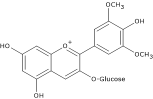
Skeletal formula of malvidin-3-glucoside, an anthocyanin
Malvidin-3-glucoside

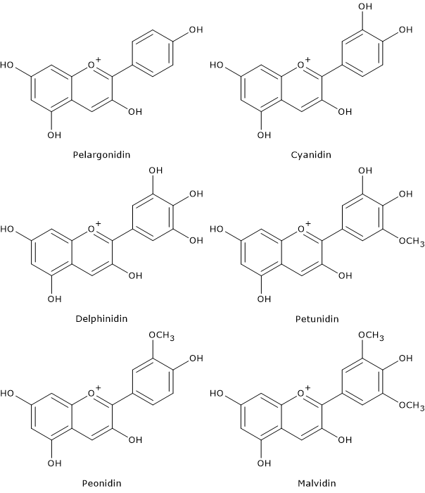
In red grapes and wine, the most abundant anthocyanins are the 3-monoglucosides of malvidin, the most abundant one both in grapes and wine, petunidin, delphinidin, peonidin, and cyanidin. In turn, the hydroxyl group at position 6 of the glucose can be acylated with an acetyl, caffeic or coumaric group, acylation that further enhances the stability.
Anthocyanidins, namely the non-conjugated molecules, are not present in grapes and in wine, except as traces.
Anthocyanins are scarcely present in white grapes and wine.
The composition of anthocyanins in wine is highly influenced both by the type of cultivar and by processing techniques, since they are present in wine as a result of extraction by maceration/fermentation processes. For this reason, wines deriving from similar varieties of grapes can have very different anthocyanin compositions.
Together with proanthocyanidins, they are the most important polyphenols in contributing to some organoleptic properties of red wine, as they are primarily responsible for astringency, bitterness, chemical stability against oxidation, as well as of the color of the young wine. In this regard, it should be underscored that with time their concentration decreases, while the color is due more and more to the formation of polymeric pigments produced by condensation of anthocyanins both among themselves and with other molecules.
During wine aging, proanthocyanidins and anthocyanins react to produce more complex molecules that can partially precipitate.

Catechins

They are, together with condensed tannins, the most abundant flavonoids, representing up to 50 percent of the total polyphenols in white grapes and between 13 percent and 30 percent in red ones.
Their levels in wine depend on the type of cultivar.

Polyphenols from grapes: skeletal formula of catechin, a flavanol
Catechin

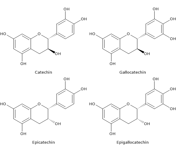
Typically, the most abundant flavanol in wine is catechin, but epicatechin and epicatechin-3-gallate are also present.

Proanthocyanidins

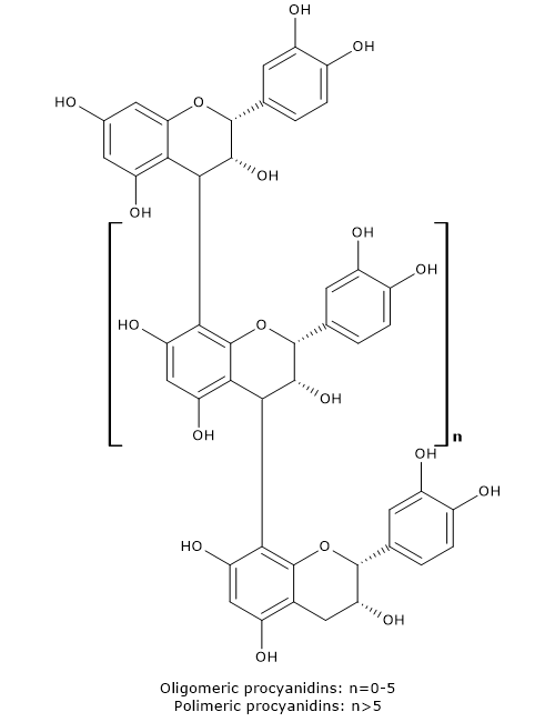
Composed of catechin monomers, they are present in the berry skin, seeds and rachis of the bunch of grapes as:

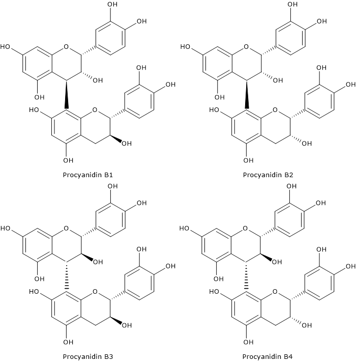
  • dimers: the most common are procyanidins B1-B4, but also procyanidins B5-B8 can be present;
  • trimers: procyanidin C1 is the most abundant;
  • tetramers;
  • polymers, containing up to 8 monomers.
Skeletal formula of procyanidin C1, a proanthocyanidin
Procyanidin C1

Their levels in wine depend on the type of grape varieties and wine-making technology, and, like anthocyanins, are much more abundant in red wines, in particular in aged wines, compared to white ones.
In addition, as previously said, together with anthocyanins, condensed tannins are important in determining some organoleptic properties of the wine.

Flavonols

Flavonols are present in a large variety of fruit and vegetables, even if in low concentrations.
They are the third most abundant group of flavonoids from grapes, after proanthocyanidins and catechins.
They are mainly present in the outer epidermis of the berry skin, where they play a role both in providing protection against UV-A and UV-B radiations and in copigmentation together with anthocyanins.
Flavanol synthesis begins in the sprout; the highest concentration is reached a few weeks after veraison, then it decreases as the berry increases in size.
Their total amount is very variable, with the red varieties often richer than the white ones.
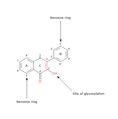
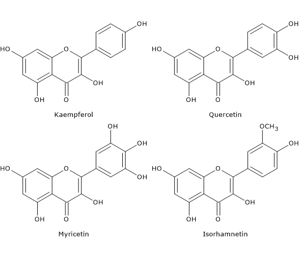
In grapes, they are present as 3-glucosides and their composition depends on the type of grapes and cultivar:

  • the derivatives of quercetin, kaempferol and isorhamnetin are found in white grapes;
  • the derivatives of myricetin, laricitrin and syringetin are found, together with the previous ones, only in red grapes, due to the lack of expression in white grapes of the gene coding for flavonoid-3′,5′-hydroxylase.
Polyphenols from grapes: skeletal formula of quercetin-3-glucoside, a flavonol
Quercetin-3-glucoside

In general, the 3-glucosides and 3-glucuronides of quercetin are the major flavonols in most of the grape varieties. Conversely, quercetin-3-rhamnoside and quercetin aglycone are the major flavonols in muscadine grapes.
In wine and grape juice, unlike grapes, they are also found as aglycones, as a result of the acid hydrolysis that occurs during processing and storage. They are present in wine in a variable amount, and the major molecules are the glycosides of quercetin and myricetin, which alone represent 20-50 percent of the total flavonols in red wine.

Hydroxycinnamic acids

Hydroxycinnamic acids are the main class of non-flavonoid polyphenols from grapes and the major polyphenols in white wine.
The most important are p-coumaric, caffeic, sinapic, and ferulic acids, present in wine as esters with tartaric acid.
They have antioxidant activity and in some white varieties of Vitis vinifera, together with flavonols, are the polyphenols mainly responsible for absorbing UV radiation in the berry.

Stilbenes

They are phytoalexins which are produced in low concentrations only by a few edible species, including grapevine (on the contrary, flavonoids are present in all higher plants).
Together with the other polyphenols from grapes and wine, also stilbenes, particularly resveratrol, have been associated with health benefits resulting from the consumption of wine.

Polyphenols from grapes: skeletal formula of trans-resveratrol, a stilbene
trans-Resveratrol

Their content increases from the veraison to the ripening of the berry, and is influenced by the type of cultivar, climate, wine-making technology, and fungal pressure.
The main stilbenes present in grapes and wine are:

  • cis- and trans-resveratrol (3,5,4′-trihydroxystilbene);
  • piceid or resveratrol-3-glucopyranoside and astringin or 3′-hydroxy-trans-piceid;
  • piceatannol;
  • dimers and oligomers of resveratrol, called viniferins, of which the most important are:

α-viniferin, a trimer;
β-viniferin, a cyclic tetramer;
γ-viniferin, a highly polymerized oligomer;
ε-viniferin, a cyclic dimer.

In grapes, other isomers and glycosylated forms of resveratrol and piceatannol, such as resveratroloside, hopeaphenol, or resveratrol di- and tri-glucoside derivatives, have been found in trace amounts.
Glycosylation of stilbenes is important for the modulation of antifungal activity, protection from oxidative degradation, and storage of the wine.
The synthesis of dimers and oligomers of resveratrol, both in grapes and wine, represents a defense mechanism against exogenous attacks or, on the contrary, the result of the action of extracellular enzymes released from pathogens in an attempt to eliminate undesirable compounds.

Hydroxybenzoates

The hydroxybenzoic acid derivatives are a minor component in grapes and wine.
In grapes, gentisic, gallic, p-hydroxybenzoic and protocatechuic acids are the main ones.

Skeletal formula of gallic acid, an hydroxybenzoic acid
Gallic Acid

Unlike hydroxycinnamates, which are present in wine as esters with tartaric acid, they are found in their free form.
Together with flavonols, proanthocyanidins, catechins, and hydroxycinnamates they are among the responsible of astringency of wine.

References

  1. Andersen Ø.M., Markham K.R. Flavonoids: chemistry, biochemistry, and applications. CRC Press Taylor & Francis Group, 2006
  2. Basli A, Soulet S., Chaher N., Mérillon J.M., Chibane M., Monti J.P.,1 and Richard T. Wine polyphenols: potential agents in neuroprotection. Oxid Med Cell Longev 2012. doi:10.1155/2012/805762
  3. de la Rosa L.A., Alvarez-Parrilla E., Gonzàlez-Aguilar G.A. Fruit and vegetable phytochemicals: chemistry, nutritional value, and stability. 1th Edition. Wiley J. & Sons, Inc., Publication, 2010
  4. Flamini R., Mattivi F., De Rosso M., Arapitsas P. and Bavaresco L. Advanced knowledge of three important classes of grape phenolics: anthocyanins, stilbenes and flavonols. Int J Mol Sci 2013;14:19651-19669. doi:10.3390/ijms141019651
  5. Georgiev V., Ananga A. and Tsolova V. Recent advances and uses of grape flavonoids as nutraceuticals. Nutrients 2014;6: 391-415. doi:10.3390/nu6010391
  6. Guilford J.M. and Pezzuto J.M. Wine and health: a review. Am J Enol Vitic 2011;62(4):471-486. doi:10.5344/ajev.2011.11013
  7. He S., Sun C. and Pan Y. Red wine polyphenols for cancer prevention. Int J Mol Sci 2008;9:842-853. doi:10.3390/ijms9050842
  8. Xia E-Q., Deng G-F., Guo Y-J. and Li H-B. Biological activities of polyphenols from grapes. Int J Mol Sci 2010;11:622-646. doi:10.3390/ijms11020622
  9. Waterhouse A.L. Wine phenolics. Ann N Y Acad Sci 2002;957:21-36. doi:10.1111/j.1749-6632.2002.tb02903.x

Polyphenols in olive oil: variability and composition

Olive oil, which is obtained from the pressing of the olives, the fruits of olive tree (Olea europaea), is the main source of lipids in the mediterranean diet, and a good source of polyphenols.
Polyphenols, natural antioxidants, are present in olive pulp and, following pressing, they pass into the oil.
Note: olives are also known as drupes or stone fruits.
The concentration of polyphenols in olive oil is the result of a complex interaction between various factors, both environmental and linked to the extraction process of the oil itself, such as:

  • the place of cultivation;
  • the cultivars (variety);
  • the level of ripeness of the olives at the time of harvesting.
    Their level usually decreases with over-ripening of the olives, although there are exceptions to this rule. For example, in warmer climates, olives produce oils richer in polyphenols, in spite of their faster maturation.
  • the climate;
  • the extraction process. In this regard, it is to underscore that the content of polyphenol in refined olive oil is not significant.

Any variation of the concentration of different polyphenols influence the taste, nutritional properties and stability of olive oil. For example, hydroxytyrosol and oleuropein (see below) give extra virgin olive oil a pungent and bitter taste.

Contents

Key polyphenols in olive oil

Among polyphenols in olive oil, there are molecules with simple structure, such as phenolic acids and alcohols, and molecules with complex structure, such as flavonoids, secoiridoids, and lignans.

Flavonoids

Flavonoids include glycosides of flavonols (rutin, also known as quercetin-3-rutinoside), flavones (luteolin-7-glucoside), and anthocyanins (glycosides of delphinidin).

Phenolic acids and phenolic alcohols

Among phenolic acids, the first polyphenols with simple structure observed in olive oil, they are found:

  • hydroxybenzoic acids, such as, gallic, protocatechuic, and 4-hydroxybenzoic acids (all with C6-C1 structure).
  • hydroxycinnamic acids, such as caffeic, vanillin, syringic, p-coumaric, and o-coumaric acids (all with C6-C3 structure).

Among phenolic alcohols, the most abundant are hydroxytyrosol, also known as 3,4-dihydroxyphenyl-ethanol, and tyrosol, also known as 2-(4-hydroxyphenyl)-ethanol.

Hydroxytyrosol

It can be present as:

  • simple phenol;
  • phenol esterified with elenolic acid, forming oleuropein and its aglycone;
  • part of the molecule verbascoside.
Hydroxytyrosol, a phenolic alcohol, and one of the polyphenols in olive oil
Hydroxytyrosol

It can also be present in different glycosidic forms, depending on the –OH group to which the glucoside, i.e. elenolic acid plus glucose, is bound.
It is one of the main polyphenols in olive oil, extra virgin olive oil, and olive vegetable water.
In nature, its concentration, such as that of tyrosol, increases during fruit ripening, in parallel with the hydrolysis of compounds with higher molecular weight, while the total content of phenolic molecules and alpha-tocopherol decreases. Therefore, it can be considered as an indicator of the degree of ripeness of the olives.
In fresh extra virgin olive oil, hydroxytyrosol is mostly present in esterified form, while in time, due to hydrolysis reactions, the non-esterified form becomes the predominant one.
Finally, the concentration of hydroxytyrosol is correlated with the stability of olive oil.

Secoiridoids

They are the polyphenols in olive oil with the more complex structure, and are produced from the secondary metabolism of terpenes.
They are glycosylated compounds and are characterized by the presence of elenolic acid in their structure (both in its aglyconic or glucosidic form). Elenolic acid is the molecule common to glycosidic secoiridoids of Oleaceae.
Unlike tocopherols, flavonoids, phenolic acids, and phenolic alcohols, that are found in many fruits and vegetables belonging to different botanical families, secoiridoids are present only in plants of the Oleaceae family.
Ligstroside, oleuropein, demethyloleuropein, and nuzenide are the main secoiridoids.
In particular, oleuropein and demethyloleuropein (as verbascoside) are abundant in the pulp, but they are also found in other parts of the fruit. Nuzenide is only present in the seeds.

Oleuropein

Oleuropein, the ester of hydroxytyrosol and elenolic acid, is the most important secoiridoid, and the main olive oil polyphenol.

<yoastmark class=

It is present in very high quantities in olive leaves, as also in all the constituent parts of the olive, including peel, pulp and kernel.
Oleuropein accumulates in olives during the growth phase, up to 14% of the net weight; when the fruit turns greener, its quantity reduces. Finally, when the olives turns dark brown, color due to the presence of anthocyanins, the reduction in its concentration becomes more evident.
It was also shown that its content is greater in green cultivars than in black ones.
During the reduction of oleuropein levels (and of the levels of other secoiridoids), an increase of compounds such as flavonoids, verbascosides, and simple phenols can be observed.
The reduction of its content is also accompanied by an increase in its secondary glycosylated products, that reach the highest values in black olives.

Lignans

Lignans, in particular (+)-1- acetoxypinoresinol and (+)-pinoresinol, are another group of polyphenols in olive oil.
(+)-pinoresinol is a common molecule in the lignin fraction of many plants, such as sesame (Sesamun indicum) and the seeds of the species Forsythia, belonging to the family Oleaceae. It has been also found in the olive kernel.
(+)-1- acetoxypinoresinol and (+)-1-hydroxypinoresinol, and their glycosides, have been found in the bark of the olive tree.

Examples of lignans, a class of pholyphenols, in olive oil
Lignans in Olive Oil

Lignans are not present in the pericarp of the olives, nor in leaves and sprigs that may accidentally be pressed with the olives.
Therefore, how they can pass into the olive oil becoming one of the main phenolic fractions is not yet known.
(+)-1- acetoxypinoresinol and (+)-pinoresinol are absent in seed oils, are virtually absent from refined virgin olive oil, while they may reach a concentration of 100 mg/kg in extra-virgin olive oil.
As seen for simple phenols and secoiridoids, there is considerable variation in their concentration among olive oils of various origin, variability probably related to differences between olive varieties, production areas, climate, and oil production techniques.

References

  1. Cicerale S., Lucas L. and Keast R. Biological activities of phenolic compounds present in virgin olive oil. Int. J. Mol. Sci. 2010;11: 458-479. doi:10.3390/ijms11020458
  2. de la Rosa L.A., Alvarez-Parrilla E., Gonzàlez-Aguilar G.A. Fruit and vegetable phytochemicals: chemistry, nutritional value, and stability. 1th Edition. Wiley J. & Sons, Inc., Publication, 2010
  3. Manach C., Scalbert A., Morand C., Rémésy C., and Jime´nez L. Polyphenols: food sources and bioavailability. Am J Clin Nutr 2004;79(5):727-747. doi:10.1093/ajcn/79.5.727
  4. Owen R.W., Mier W., Giacosa A., Hull W.E., Spiegelhalder B. and Bartsch H. Identification of lignans as major components in the phenolic fraction. Clin Chem 2000;46:976-988.
  5. Tripoli E., Giammanco M., Tabacchi G., Di Majo D., Giammanco S. and La Guardia M. The phenolic compounds of olive oil: structure, biological activity and beneficial effects on human health. Nutr Res Rev 2005:18;98-112. doi:10.1079/NRR200495

Black tea: processing, properties, and health benefits

Black tea, like the other types of tea, is an infusion of dried and processed leaves of Camellia sinensis, a shrub belonging to the Theaceae family.
Unlike what happens during green tea production, during black tea production the almost complete oxidation of the substances contained in the leaves occurs, particularly catechins, polyphenols of the flavonoid group.
The color of the processed leaves is dark, whereas the beverage is brown-red in color.
Black tea is prepared with one tea bag per person, or one teaspoon per person in case of loose tea leaves, with an infusion time of 3-4 minutes in water at 95-100 °C.
Tea is a beverage with ancient origins, dating back to almost 4,000 years ago. It is one of the most-consumed beverages, particularly in Asia, where the favorite tea is green tea, especially in Japan and China, whereas black tea is preferred by Western populations and at global level, accounting for about 78 percent of the tea consumed.
The oxidation of the compounds present in the leaves during processing reduces the potential beneficial effects ascribed to the polyphenols initially present.

Contents

How black tea is made

All the types of teas are produced from fresh leaves of Camellia sinensis. During harvesting, young leaves are preferred, as the older ones are considered inferior in quality.
The processing that leads to the production of loose dried tea leaves ready for brewing black tea proceeds through four steps: withering, rolling, oxidation, and drying. Such processing leads to the near complete oxidation of the substances present, particularly catechins.
Withering is the process by which the water present in the leaves, about 75 percent of the leaf’s weight, is removed, thus causing sap concentration. Withering, which makes the next step easier, can be achieved in three different ways:

  • by exposing leaves to sunlight;
  • by appropriately heating the rooms where the leaves are stored;
  • by machineries that ventilate the leaves.

The rolling step follows the withering of the leaves, and, breaking the leaf tissues, facilitates the outflow of lymph thus promoting the subsequent oxidation of polyphenols.
The oxidation step is also improperly called fermentation. In this step, the oxidation by atmospheric oxygen and polyphenol oxidase (EC 1.10.3.1) of 90-95 percent of the polyphenols occurs, accompanied by other changes that color the leaves with a red color. Temperature, typically 25°C, pH, relative humidity, 95 percent or more, ventilation, and duration are crucial factors, too. This step is crucial in determining the quality of the tea, as it gives it its organoleptic characteristics and composition in polyphenols, quite different from those of green tea, which is produced in such a way as to minimize oxidation processes.
Note that caffeine content does not vary significantly.
Drying is the last step. It is carried out at a temperature of 80-90 °C for about 20-25 minutes. The high temperature inactivates polyphenol oxidase, and then stops enzymatic oxidation processes.

Thearubigins and theaflavins

The oxidative processes that occur during black tea production affect monomeric and gallate catechins, to a lesser extent catechins glycosides, especially myricetin, and non-flavonoid compounds, such as teagallin, and leads to the formation of complex polyphenols such as thearubigins, theaflavins and theaflavic acids.
Thearubigins, brownish in color, are polymers of catechins not yet well characterized and the major polyphenols in black tea, accounting for about 20 percent of the dry leaf weight. They contribute to the richness in taste and color.
Theaflavins, red-orange in color, are dimers of catechins and account for about 3-5 percent of the dry leaf weight. They contribute to the astringent and brisk taste, as well as the red-orange in color.
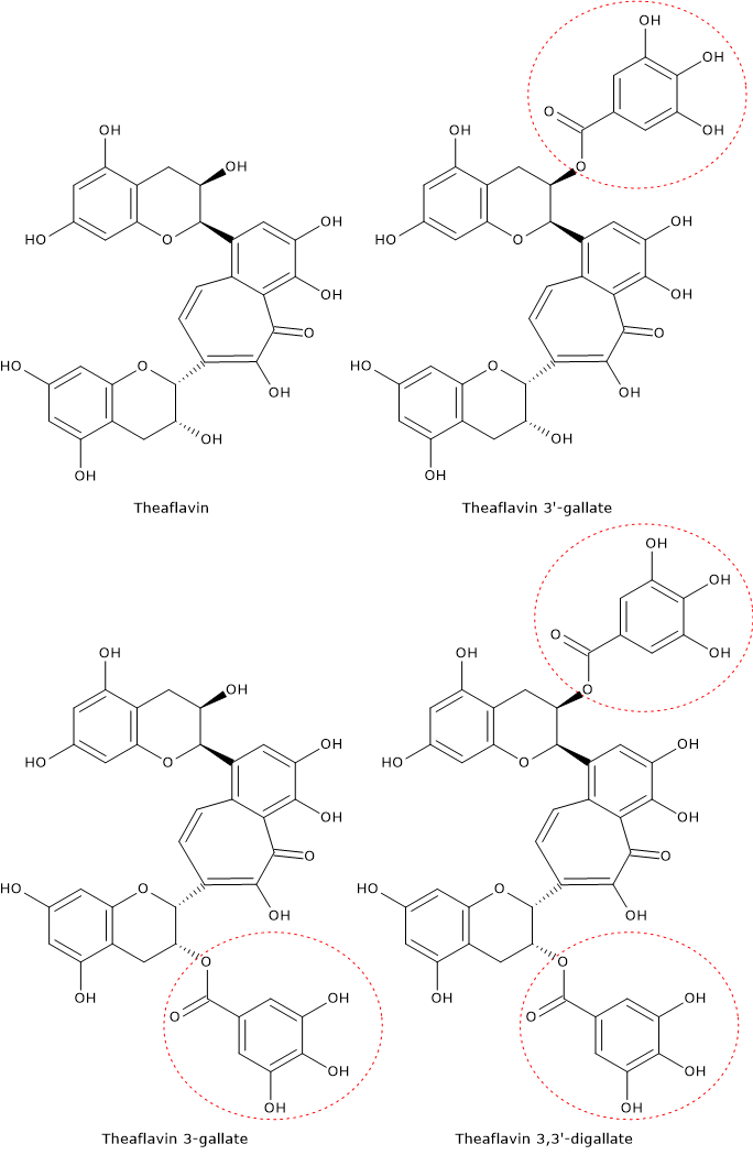
The main theaflavins are:

  • theaflavin 3-gallate;
  • theaflavin 3′-gallate;
  • theaflavin 3,3’-digallate, the most abundant.
Skeletal formulas of theaflavins, dimers of catechins present in black tea
Theaflavins

Health benefits

The health benefits of black tea are largely due to its complex polyphenols, thearubigins and theaflavins, being catechins largely oxidized during leaf processing.
Here are three examples.

  • Theaflavins have been highlighted as having antiviral activity which, similarly to catechins, appears to be particularly effective against positive single-stranded RNA viruses. These viruses also include SARS-CoV-1 and SARS-CoV-2, viruses belonging to the Coronaviridae family.
    Like catechins, the galloyl groups appear to be important for the antiviral activity of theaflavins.
  • The phytochemicals present in black tea, like those in green tea, seem to be able to reduce the glycemic index of starchy foods. The effect appears to be due to the inhibition of the activity of pancreatic alpha-amylase and other digestive enzymes, and to the direct interaction with starch, that would reduce the surface available to enzyme activity. The inhibition is greater on gluten free foods; this seems to be due to the action of gluten on complex polyphenols that would not be able to interact with the polysaccharide. For more information, see the article on tea polyphenols.
  • Thearubigins and theaflavins seem to have anticariogenic effects due to the inhibitory action on salivary and bacterial amylase, and seem to be more effective than green tea catechins.
    Moreover, black tea seems to be able to inhibit acid production in the oral cavity.

References

  1. Asil M.H., Rabiei B., Ansari R.H. Optimal fermentation time and temperature to improve biochemical composition and sensory characteristics of black tea. Aust J Crop Sci 2012;6(3):550-8.
  2. Kan L., Capuano E., Fogliano V., Oliviero T. and Verkerk R. Tea polyphenols as a strategy to control starch digestion in bread: the effects of polyphenol type and gluten. Food Funct 2020;11:5933-5943. doi:10.1039/D0FO01145B
  3. Kuhnert N. Unraveling the structure of the black tea thearubigins. Arch Biochem Biophys 2010;501(1):37-51. doi:10.1016/j.abb.2010.04.013
  4. Li S., Lo C-Y., Pan M-H., Lai C-S. and Ho C-T. Black tea: chemical analysis and stability. Food Funct 2013;4:10-18. doi:10.1039/C2FO30093A
  5. Mhatre S., Srivastava T., Naik S., Patravale V. Antiviral activity of green tea and black tea polyphenols in prophylaxis and treatment of COVID-19: a review. Phytomedicine 2020;153286. doi:10.1016/j.phymed.2020.153286
  6. Menet M-C., Sang S., Yang C.S., Ho C-T., and Rosen R.T. Analysis of theaflavins and thearubigins from black tea extract by MALDI-TOF mass spectrometry. J Agric Food Chem 2004;52:2455-61. doi:10.1021/jf035427e
  7. Sharma V.K., Bhattacharya A., Kumar A. and Sharma H.K. Health benefits of tea consumption. Trop J Pharm Res 2007;6(3):785-792.

Green tea: processing, properties, and health benefits

Green tea, like the other types of tea, is an infusion of dried and processed leaves of Camellia sinensis, a member of the Theaceae family.
The processing of the leaves leading to the product ready for use is such as to minimize the oxidation of the compounds contained in them, particularly phytochemicals such as catechins, which are polyphenols belonging to the class of flavonoids and the most responsible for the health benefits of green tea.
Having undergone no significant chemical modifications, leaves retain green color, whereas the beverage, prepared with one tea bag per person, or in case of loose tea, one teaspoon per person, for an infusion time of about 3 minutes in water at 75 °C, is golden yellow in color.
Some organoleptic properties of green tea, such as the flavor, that is more delicate and lighter than that of black tea, and the health properties, which have always been recognized in East Asia cultures, depend on leaf processing.
Only recently scientists started studying the health benefits of tea consumption, highlighting its role in preventing many diseases, such as cardiovascular diseases and some types of cancer.
It has been shown that tea polyphenols, particularly catechins, are able to activate intracellular signaling pathways by binding to membrane receptors and/or entering the cell and binding to cytoplasmic, mitochondrial or nuclear receptors. Then, depending on the cell type, they activate or inhibit some cellular processes.
Given the high consumption of tea in the world, even small effects on health could have significant effects on public health.

Contents

Camellia sinensis

Camellia sinensis is an evergreen plant native to South, East, and Southeast Asia, which is now cultivated in at least 30 countries, mostly in tropical or subtropical climates, although some varieties grow in Cornwall and Washington State.
In nature, Camellia sinensis can grow up to 15-20 meters (49-65 ft), whereas in plantations it is pruned to less than 1,5 meters to facilitate leaf harvesting.
It can grow up to altitude of 1,500-2,000 meters (4,900-6,550 ft), and many of the high-quality teas are produced from such crops, as the plant grows slowly and the leaves acquire a better flavor.
The most cultivated varieties, of the four known, are Camellia sinensis var. sinensis, native to China, and Camellia sinensis var. assamica, native to India.
The different types of tea are produced from fresh leaves. Young leaves are preferred over older leaves that are considered to be inferior in quality.
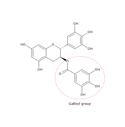
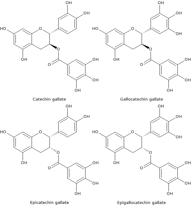
Fresh leaves are rich in water-soluble polyphenols, especially catechins and glycosylated catechins. The major catechins in green tea are epigallocatechin-3-gallate or EGCG, the most active, epigallocatechin, epicatechin 3-gallate, epicatechin. Catechin, gallocatechin, catechin gallate, and gallocatechin gallate are also present, although in lower amount.

Skeletal formula of gallocatechin gallate, one of the catechins found in green tea
Gallocatechin gallate

These polyphenols account for 30-42 percent of the dry leaf weight. Caffeine accounts for 1,5-4,5 percent of the dry leaf weight.
In addition to leaf processing, the organoleptic properties of the beverage are influenced by cultivar, characteristics of the soil where the plant grown up, methods of cultivation, altitude, climate, and time of year in which leaf harvest occurs.

How green tea is made

The differences in leaf processing, which lead to the different types of tea ready for consumption, cause different degrees of oxidation of the compounds present in them, especially catechins.
During green tea manufacturing, oxidative processes, both enzymatic and chemical, are minimized. After harvesting, leaves are exposed to sunlight for 2-3 hours and withered/dried. Then, the processing proceeds through three steps:

  • heat treatment;
  • rolling;
  • drying.

Heat treatment, short and gentle, is crucial for the quality and properties of the beverage. It can done either with a steam, the traditional Japanese method, or by dry cooking in hot pans, that is similar to a roasting method and is the traditional Chinese method. Heat treatment inactivates enzymes and then prevents the enzymatic oxidation processes, particularly those involving polyphenols. It also removes the grassy smell, and evaporates, in the case of the traditional Chinese method, part of the water of the leaf, which constitutes about 75 percent of its weight, making it softer, thus facilitating the next step.
Heat treatment is followed by the rolling step, that facilitates the subsequent drying step and, destroying the leaf tissue, favors the release of aromas, thus improving the quality of the product.
The drying, the last step, improves the appearance of beverage and leads to the production of new compounds.

Health benefits

In East Asia cultures, mainly in China and Japan, tea drinking has always been associated with health benefits. Below is a brief review of the results of epidemiological and laboratory studies that have analyzed the effects that green tea consumption can play in preventing many diseases. EGCG, which is the most abundant catechin in green tea accounting for about 60 percent of the polyphenols present in dried leaves, seems to play the main role.
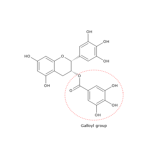
At the molecular level, the galloyl groups at positions 3 and/or 3′ appear to be essential for many of the effects exerted by catechins.

Cardiovascular disease

Cardiovascular disease is the main cause of deaths worldwide, particularly in low- and middle-income countries, with an estimate of about 17 million deaths in 2008 that could increase up to 23.3 million by 2030.
Daily tea consumption, especially green tea, seems to be associated with a reduced risk of developing cardiovascular disease, such as hypertension and stroke.
Among the proposed mechanisms, the improved bioactivity of the endothelium-derived vasodilator nitric oxide, due to the action of tea polyphenols that could enhance nitric oxide synthesis and/or decrease its breakdown by superoxide anions, seem to be important.

Cancer

Several epidemiological and laboratory studies have shown encouraging results with respect to the preventive role of tea consumption, especially green tea, against the development of some cancers such as those of the oral cavity, digestive tract, and lung among those who have never smoked.
Tea polyphenols seem to act not only as antioxidants, but also as compounds that, directly, can influence gene expression and various metabolic pathways.

Antiviral activity

Recent studies have highlighted antiviral effects of catechins, particularly EGCG of green tea and theaflavins of black tea, especially against positive single-stranded RNA viruses, to which the Coronaviridae family, and then SARS-CoV-1 and SARS-CoV-2, belongs.
The antiviral properties of EGCG appear to be due to its structural characteristics, namely, the presence of pyrogallic and galloyl groups.

Starch digestion

In vitro studies have shown that green tea and black tea polyphenols can reduce the glycemic index of starchy foods. Hence, they could be useful in controlling their glycemic index in vivo. This effect seems to be due to the inhibition of pancreatic alpha-amylase and other digestive enzymes, and to a direct interaction between starch and phytochemicals that would reduce the surface area of starch granules available for enzyme activity. Green tea appears to be equally effective against both gluten-containing foods, against which black tea appears less effective, and gluten free foods.

Weight loss

During weight loss and weight-loss maintenance it is important to keep as constant as possible the daily energy expenditure.
Since the 90s, it has been proposed that green tea, by virtue of its content of caffeine and catechins could be of help for:

  • maintaining, or even increasing, daily energy expenditure;
  • increasing fat oxidation.

In addition to these potential lipolytic and thermogenic effects, catechins and caffeine could act on other targets, such as lipid absorption and energy intake, perhaps through their effect on gut microbiota, which is part of the larger human microbiota, and gene expression.
And products for weight loss and weight maintenance based on green tea extracts have been marketed. It should be noted that these products contain catechins and caffeine in much higher amount than beverage.
How much truth is there in green tea’s fat burning effect?
The issue seems to have been resolved by a meta-analysis of 15 studies on weight loss and intake of these products. The study showed that green tea-based products induces, in overweight and obese adults, a weight loss that is:

  • not statistically significant;
  • very small;
  • probably not clinically important.

Hence, on the basis of scientific evidence, green tea does not seem to be helpful in fat loss nor in weight maintenance.

Anticariogenic activity

Animal and in vitro studies have shown that tea, and particularly its polyphenols, seem to possess:

  • antibacterial activity against cariogenic bacteria, such as Streptococcus mutans, as in the case of green tea EGCG;
  • inhibitory action on salivary and bacterial amylase, in which black tea thearubigins and theaflavins are more effective than green tea catechins;
  • inhibitory action on acid production in the oral cavity.

All these properties make green tea and black tea beverages with potential anticariogenic activity.

References

  1. Arab L., Khan F., and Lam H. Tea consumption and cardiovascular disease risk. Am J Clin Nutr 2013;98:1651S-1659S. doi:10.3945/ajcn.113.059345
  2. Clifford M.N., van der Hooft J.J.J., and Crozier A. Human studies on the absorption, distribution, metabolism, and excretion of tea polyphenols. Am J Clin Nutr 2013;98:1619S-1630S. doi:10.3945/ajcn.113.058958
  3. Dwyer J.T. and Peterson J. Tea and flavonoids: where we are, where to go next. Am J Clin Nutr 2013;98:1611S-1618S. doi:10.3945/ajcn.113.059584
  4. Goenka P., Sarawgi A., Karun V., Nigam A.G., Dutta S., Marwah N. Camellia sinensis (Tea): implications and role in preventing dental decay. Phcog Rev 2013;7:152-156. doi:10.4103/0973-7847.120515
  5. Green R.J., Murphy A.S., Schulz B., Watkins B.A. and Ferruzzi M.G. Common tea formulations modulate in vitro digestive recovery of green tea catechins. Mol Nutr Food Res 2007;51(9):1152-1162. doi:10.1002/mnfr.200700086
  6. Grassi D., Desideri G., Di Giosia P., De Feo M., Fellini E., Cheli P., Ferri L., and Ferri C. Tea, flavonoids, and cardiovascular health: endothelial protection. Am J Clin Nutr 2013;98:1660S-1666S. doi:10.3945/ajcn.113.058313
  7. Huang W-Y., Lin Y-R., Ho R-F., Liu H-Y., and Lin Y-S. Effects of water solutions on extracting green tea leaves. Sci World J 2013;Article ID 368350. doi:10.1155/2013/368350
  8. Hursel R. and Westerterp-Plantenga M.S. Catechin- and caffeine-rich teas for control of body weight in humans. Am J Clin Nutr 2013;98:1682S-1693S. doi:10.3945/ajcn.113.058396
  9. Hursel R., Viechtbauer W. and Westerterp-Plantenga M.S. The effects of green tea on weight loss and weight maintenance: a meta-analysis. Int J Obesity 2009;33:956-961. doi:10.1038/ijo.2009.135
  10. Jurgens T.M., Whelan A.M., Killian L., Doucette S., Kirk S., Foy E. Green tea for weight loss and weight maintenance in overweight or obese adults. Editorial group: Cochrane Metabolic and Endocrine Disorders Group. 2012:12 Art. No.: CD008650. doi:10.1002/14651858.CD008650.pub2
  11. Lambert J.D. Does tea prevent cancer? Evidence from laboratory and human intervention studies. Am J Clin Nutr 2013;98:1667S-1675S. doi:10.3945/ajcn.113.059352
  12. Lorenz M. Cellular targets for the beneficial actions of tea polyphenols. Am J Clin Nutr 2013;98:1642S-1650S. doi:10.3945/ajcn.113.058230
  13. Mathur A., Gopalakrishnan D., Mehta V., Rizwan S.A., Shetiya S.H., Bagwe S. Efficacy of green tea-based mouthwashes on dental plaque and gingival inflammation: a systematic review and meta-analysis. Indian J Dent Res 2018;29(2):225-232. doi:10.4103/ijdr.IJDR_493_17
  14. Mhatre S., Srivastava T., Naik S., Patravale V. Antiviral activity of green tea and black tea polyphenols in prophylaxis and treatment of COVID-19: a review. Phytomedicine 2020;153286. doi:10.1016/j.phymed.2020.153286
  15. Sharma V.K., Bhattacharya A., Kumar A. and Sharma H.K. Health benefits of tea consumption. Trop J Pharm Res 2007;6(3):785-792.
  16. Yang Y-C., Lu F-H., Wu J-S., Wu C-H., Chang C-J. The protective effect of habitual tea consumption on hypertension. Arch Intern Med 2004;164:1534-1540. doi:10.1001/archinte.164.14.1534
  17. Yuan J-M. Cancer prevention by green tea: evidence from epidemiologic studies. Am J Clin Nutr 2013;98:1676S-1681S. doi:10.3945/ajcn.113.058271
  18. Xu J., Xu Z., Zheng W. A review of the antiviral role of green tea catechins. Molecules 2017;22(8):1337. doi:10.3390/molecules22081337

Anthocyanins: food sources, absorption, and metabolism

Together with catechins and proanthocyanidins, anthocyanins and their oxidation products are the most abundant flavonoids in the human diet.
Examples of anthocyanin rich foods are:

  • certain varieties of grains, such as some types of pigmented rice (e.g. black rice) and maize (purple corn);
  • in certain varieties of root and leafy vegetables such as aubergine, red cabbage, red onions and radishes, beans;
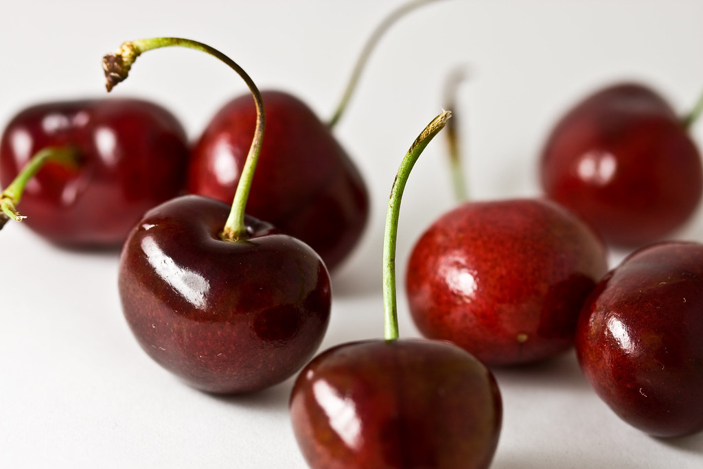
  • but especially in red fruits.

Example of anthocyanin rich food
Anthocyanins are also present in red wine; as the wine ages, they are transformed into various complex molecules.
Anthocyanin content in vegetables and fruits is generally proportional to their color: it increases during maturation, and it reaches values up to 4 g/kg fresh weight (FW) in cranberries and black currants.
These polyphenols are found primarily in the skin, except for some red fruits, such as cherries and red berries (e.g. strawberries), in which they are present both in the skin and flesh.
Glycosides of cyanidin are the most common anthocyanins in foods.

Contents

Anthocyanin rich fruits

  • Berries are the main source of anthocyanins, with values ranging between 67 and 950 mg/100 g FW.
  • Other fruits, such as red grapes, cherries and plums, have content ranging between 2 and 150 mg/100 g FW.
  • Finally, in fruits such as nectarines, peaches, and some types of apples and pears, anthocyanins are poorly present, with a content of less than 10 mg/100 g FW.

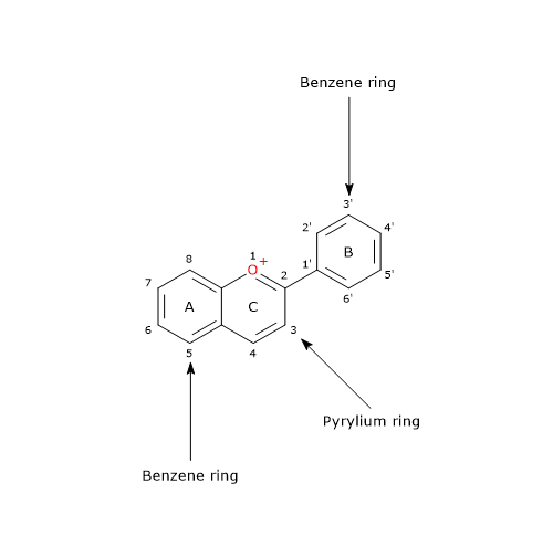
Cranberries, besides their very high content of anthocyanins, are one of the rare food that contain glycosides of the six most commonly anthocyanidins present in foods: pelargonidin, delphinidin, cyanidin, petunidin, peonidin, and malvidin. The main anthocyanins are the 3-O-arabinosides and 3-O-galactosides of peonidin and cyanidin. A total of 13 anthocyanins have been detected, mainly 3-O-monoglycosides.

Anthocyanin absorption

Until recently, it was believed that anthocyanins, together with proanthocyanidins and gallic acid ester derivatives of catechins, were the least well-absorbed polyphenols, with a time of appearance in the plasma consistent with the absorption in the stomach and small intestine. Indeed, some studies have shown that their bioavailability has been underestimated since, probably, all of their metabolites have not been yet identified.
In this regard, it should be underlined that only a small part of the food anthocyanins is absorbed in their glycated forms or as hydrolysis products in which the sugar moiety has been removed. Therefore, a large amount of these ingested polyphenols enters the colon, where they can also suffer methylation, sulphatation, glucuronidation and oxidation reactions.

Anthocyanins and colonic microbiota

Few studies have examined the metabolism of anthocyanins by the gut microbiota in the colon, which is part of the larger human microbiota.
Within two hours, it seems that all the anthocyanins lose their sugar moieties, thus producing anthocyanidins.
Anthocyanidins are chemically unstable in the neutral pH of the colon. They can be metabolized by colonic microbiota or chemically degraded producing a set of new molecules that have not yet fully identified, but which include phenolic acids such as gallic acid, syringic acid, protocatechuic acid, vanillic acid and phloroglucinol or 1,3,5-trihydroxybenzene. These molecules, thanks to their higher microbial and chemical stability, might be the main responsible for the antioxidant activities and the other physiological effects that have been observed in vivo and attributed to anthocyanins.

References

  1. de la Rosa L.A., Alvarez-Parrilla E., Gonzàlez-Aguilar G.A. Fruit and vegetable phytochemicals: chemistry, nutritional value, and stability. 1th Edition. Wiley J. & Sons, Inc., Publication, 2010
  2. de Pascual-Teresa S., Moreno D.A. and García-Viguera C. Flavanols and anthocyanins in cardiovascular health: a review of current evidence. Int J Mol Sci 2010;11:1679-1703. doi:10.3390/ijms11041679
  3. Escribano-Bailòn M.T., Santos-Buelga C., Rivas-Gonzalo J.C. Anthocyanins in cereals. J Chromatogr A 2004:1054;129-141. doi:10.1016/j.chroma.2004.08.152
  4. Han X., Shen T. and Lou H. Dietary polyphenols and their biological significance. Int J Mol Sci 2007;9:950-988. doi:10.3390/i8090950
  5. Manach C., Scalbert A., Morand C., Rémésy C., and Jime´nez L. Polyphenols: food sources and bioavailability. Am J Clin Nutr 2004;79(5):727-747. doi:10.1093/ajcn/79.5.727
  6. Tsao R. Chemistry and biochemistry of dietary polyphenols. Nutrients 2010;2:1231-1246. doi:10.3390/nu2121231

Tea polyphenols: preventive effects and mechanism of actions

The leaves of the tea plant, Camellia sinensis, are rich in compounds with many biological activities, ranging from preventing the development of chronic diseases to reducing the glycemic index of starchy foods.
More than 4000 different molecules have been found in the beverage, of which about one third of these are polyphenols, the most important phytochemicals in determining the nutritional values and health benefits of the tea.
Tea polyphenols are mostly flavonoids. Examples are catechins in green tea, among which epigallocatechin-3-gallate or EGCG is the most important and abundant, and thearubigins and theaflavins in black tea. Their galloyl groups at positions 3 and/or 3′ appear to be particularly important for their effects.

Skeletal formula of epigallocatechin gallate, a catechin, and one of the tea polyphenols
Epigallocatechin Gallate

Other bioactive compounds present in tea leaves are:

  • alkaloids, such as caffeine, theophylline and theobromine;
  • amino acids, and among them, theanine or R-glutamylethylamide, that is also a brain neurotransmitter and one of the most important amino acids in green tea;
  • proteins;
  • carbohydrates;
  • chlorophyll;
  • volatile organic molecules, that contribute to the aroma of the beverage;
  • fluoride, aluminum and trace elements.

Contents

Biological activities

Polyphenols, both in vivo and in vitro, have a broad spectrum of biological activities, such as:

  • antioxidant and prooxidant properties;
  • a protective role against the development of diabetes, hyperlipidemia, and various types of tumors;
  • inhibition of inflammation;
  • antiviral activities;
  • anticariogenic activity.

Hence, there has been a growing interest in recent years toward the possible preventive effects of tea against many diseases, particularly cardiovascular disease, for example in the development and progression of atherosclerosis.

Mechanisms of action

Currently, knowledge is accumulating on the effects of tea polyphenols at cellular and molecular level.
It seems, at least in vitro, that catechins, and theaflavins and thearubigins are the compounds responsible for the physiological effects and health benefits of green tea and black tea, respectively.
Among the molecular mechanisms by which tea polyphenols seem to exert their effects, it has been observed, after binding to specific cell membrane receptors, a change in the activity of various protein kinases that then phosphorylate target proteins, such as transcription factors, that, in turn, translocate into the nucleus and modify the gene expression. This appears to be the mechanism of action of EGCG, and the mechanism proposed for thearubigins, polymeric polyphenols that, due to their large dimensions, may not be able to cross the plasma membrane.
In addition, some polyphenols could be able to cross the plasma membrane, then binding to specific cytoplasmic, mitochondrial or nuclear targets.
And, depending on the cell type and their amount, tea polyphenols can activate or inhibit certain cellular processes.

Starch digestion

Tea polyphenols exert an inhibitory effect on starch digestion.
In vitro studies have shown that green tea extracts, which contain monomeric polyphenols, have an equal inhibitory effect on starch digestibility of wheat bread and gluten free bread, whereas black tea extracts, rich in tannins, namely, polymeric polyphenols, are less effective against wheat bread. Therefore, it seems that the inhibitory effect of tannins is negatively influenced by gluten, whereas gluten has a lower inhibitory effect on monomeric polyphenols.
The inhibitory effect of these phytochemicals has been attributed to various molecular mechanisms briefly described below.

  • A competitive inhibition on pancreatic alpha-amylase. The galloyl groups are thought to be important for this effect.
  • The inhibition of other digestive enzymes present in the gastrointestinal tract.
  • The direct interaction with starch. Tea polyphenols can interact with starch granules through hydrogen bonds and hydrophobic forces, thus reducing the available surface to react with digestive enzymes.
  • Conversely, gluten could reduce the amount of polyphenols able to interact with starch and therefore able to inhibit its digestion.

Tea polyphenols could represent a means for controlling the glycemic index of starchy foods. However, it should be emphasized that, for example in the case of bread, to achieve an inhibitory effect, 100 g of bread must be co-digested with 2.5 cups of green tea or 2 cups of black tea.

References

  1. Arab L., Khan F., and Lam H. Tea consumption and cardiovascular disease risk. Am J Clin Nutr 2013;98:1651S-1659S. doi:10.3945/ajcn.113.059345
  2. Dwyer J.T. and Peterson J. Tea and flavonoids: where we are, where to go next. Am J Clin Nutr 2013;98:1611S-1618S. doi:10.3945/ajcn.113.059584
  3. Grassi D., Desideri G., Di Giosia P., De Feo M., Fellini E., Cheli P., Ferri L., and Ferri C. Tea, flavonoids, and cardiovascular health: endothelial protection. Am J Clin Nutr 2013;98:1660S-1666S. doi:10.3945/ajcn.113.058313
  4. Kan L., Capuano E., Fogliano V., Oliviero T. and Verkerk R. Tea polyphenols as a strategy to control starch digestion in bread: the effects of polyphenol type and gluten. Food Funct 2020;11:5933-5943. doi: 10.1039/D0FO01145B
  5. Lambert J.D. Does tea prevent cancer? Evidence from laboratory and human intervention studies. Am J Clin Nutr 2013;98:1667S-1675S. doi:10.3945/ajcn.113.059352
  6. Lenore Arab L., Khan F., and Lam H. Tea consumption and cardiovascular disease risk. Am J Clin Nutr 2013;98:1651S-1659S. doi:10.3945/ajcn.113.059345
  7. Lorenz M. Cellular targets for the beneficial actions of tea polyphenols. Am J Clin Nutr 2013;98:1642S-1650S. doi:10.3945/ajcn.113.058230
  8. Mathur A., Gopalakrishnan D., Mehta V., Rizwan S.A., Shetiya S.H., Bagwe S. Efficacy of green tea-based mouthwashes on dental plaque and gingival inflammation: a systematic review and meta-analysis. Indian J Dent Res 2018;29(2):225-232. doi:10.4103/ijdr.IJDR_493_17
  9. Mhatre S., Srivastava T., Naik S., Patravale V. Antiviral activity of green tea and black tea polyphenols in prophylaxis and treatment of COVID-19: a review. Phytomedicine 2020;153286. doi:10.1016/j.phymed.2020.153286
  10. Sharma V.K., Bhattacharya A., Kumar A. and Sharma H.K. Health benefits of tea consumption. Trop J Pharm Res 2007;6(3):785-792.
  11. Yuan J-M. Cancer prevention by green tea: evidence from epidemiologic studies. Am J Clin Nutr 2013;98:1676S-1681S. doi:10.3945/ajcn.113.058271
  12. Xu J., Xu Z., Zheng W. A review of the antiviral role of green tea catechins. Molecules 2017;22(8):1337. doi:10.3390/molecules22081337

Isoflavones: structure, foods and health effects

Isoflavones are colorless polyphenols belonging to the flavonoid family.
Unlike the majority of the other flavonoids, they have a restricted taxonomic distribution, being present almost exclusively in the Leguminosae or Fabaceae plant family, mainly in soy.
Since legumes, soy in primis, are a major part of the diet in many cultures, these flavonoids may have a great impact on human health.
They are also present in beans and broad beans, but in much lower concentrations than those found in soy and soy products.
Also red clover or meadow clover (Trifolium pratense), another member of Leguminosae family, is a good source.
Currently, they are not found in fruits and vegetables.
Together with phenolic acids, such as caffeic acid and gallic acid, and quercetin glycosides, they are the most well-absorbed polyphenols, followed by flavanones and catechins,but not gallocatechins.
In plants, some isoflavones have antimicrobial activity and are synthesized in response to attacks by bacteria or fungi; thus they act as phytoalexins.

Contents

Chemical structure

While most flavonoids have B ring attached to position 2 of C ring, isoflavones have B ring attached to position 3 of C ring.

Basic skeleton structure of isoflavones, polyphenols belonging to the flavonoid family
Basic Skeleton of Isoflavones

Even if they are not steroids, they have structural similarities to estrogens, particularly estradiol. This confers them pseudohormonal properties, such as the ability to bind estrogen receptors; therefore, they are classified as phytoestrogens or plant estrogens. The benefits often ascribed to soy and soy products, for example tofu, are believed to result from the ability of isoflavones to act as estrogen mimics .
It should be underlined that the binding to estrogen receptors seems to lose strength with time, therefore their potential efficacy should not be overestimated.
In foods, they are present in four forms:

  • aglycone;
  • 7-O-glucoside;
  • 6′-O-acetyl-7-O-glucoside;
  • 6′-O-malonyl-7-O-glucoside.

Soy isoflavones: genistein, daidzein and glycitein

Soy and soy products, such as soy milk, tofu, tempeh and miso, are the main source of isoflavones in the human diet.
The isoflavone content of soy and soy products varies greatly as a function of growing conditions, geographic zone, and processing; for example, in soy it ranges between 580 and 3800mg/kg fresh weight, while in soy milk it range between 30 and 175 mg/L. The most abundant isoflavones in soy and soy products are genistein, daidzein and glycitein, generally present in a concentration ratio of 1:1:0,2. Biochanin A and formononetin are other isoflavones present in less concentrations.

Basic skeleton structure of isoflavones
Isoflavones

The 6′-O-malonyl derivatives have a bitter, unpleasant, and astringent taste; therefore they give a bad flavor to the food in which they are contained. However, being sensitive to temperature, they are often hydrolyzed to glycosides during processing, such as the production of soy milk.
The fermentation processes needed for the preparation of certain foods, such as tempeh and miso, determines in turn the hydrolysis of glycosides to aglycones, i.e. the sugar-free molecule.
Isoflavone glycosides present in soy and soy products can also be deglycosylated by β-glucosidases in the small intestine.
The aglycones are very resistant to heat.
Although many compounds present in the diet are converted by intestinal bacteria to less active molecules, other compounds are converted to molecules with increased biological activity. This is the case of isoflavones, but also of prenylflavonoids from hops (Humulus lupulus), and lignans, that are other phytoestrogens.

Phytoestrogens and menopause

Vasomotor symptoms, such as night sweats and hot flashes, and bone loss are very common in perimenopause, also called menopausal transition, and menopause. Hormone replacement therapy (HRT) has proved to be a highly effective treatment for the prevention of menopausal bone loss and vasomotor symptoms.
The use of alternative therapies based on phytoestrogens is increased as a result of the publication of the “Women’s Health Initiative” study, that suggests that hormone replacement therapy could lead to more risks than benefits, in particular an increased risk of developing some chronic diseases.
Soy isoflavones are among the most used phytoestrogens by menopausal women, often taken in the form of isoflavone fortified foods or isoflavone supplements.
However, many studies have highlighted the lack of efficacy of soy isoflavones, and red clover isoflavones, even in large doses, in the prevention of vasomotor symptoms, such as hot flushes and night sweats, and bone loss during menopause.

References

  1. de la Rosa L.A., Alvarez-Parrilla E., Gonzàlez-Aguilar G.A. Fruit and vegetable phytochemicals: chemistry, nutritional value, and stability. 1th Edition. Wiley J. & Sons, Inc., Publication, 2010
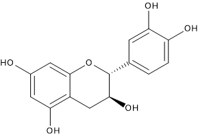
  2. Han X., Shen T. and Lou H. Dietary polyphenols and their biological significance. Int J Mol Sci 2007;9:950-988. doi:10.3390/i8090950
  3. Lagari V.S., Levis S. Phytoestrogens for menopausal bone loss and climacteric symptoms. J Steroid Biochem Mol Biol 2014;139:294-301 doi:10.1016/j.jsbmb.2012.12.002
  4. Lethaby A., Marjoribanks J., Kronenberg F., Roberts H., Eden J., Brown J. Phytoestrogens for menopausal vasomotor symptom. Cochrane Database of Systematic Reviews 2013, Issue 12. Art. No.: CD001395. doi:10.1002/14651858.CD001395.pub4
  5. Levis S., Strickman-Stein N., Ganjei-Azar P., Xu P., Doerge D.R., Krischer J. Soy isoflavones in the prevention of menopausal bone loss and menopausal symptoms: a randomized, double-blind trial. Arch Intern Med 2011:8;171(15):1363-1369 doi:10.1001/archinternmed.2011.330
  6. Manach C., Scalbert A., Morand C., Rémésy C., and Jime´nez L. Polyphenols: food sources and bioavailability. Am J Clin Nutr 2004;79(5):727-747. doi:10.1093/ajcn/79.5.727
  7. Tsao R. Chemistry and biochemistry of dietary polyphenols. Nutrients 2010;2:1231-1246. doi:10.3390/nu2121231

Procyanidins: food sources and health benefits

The interest on proanthocyanidins, and their content in foods, has increased as a result of the discovery, due to clinical and laboratory studies, of their anti-infectious, anti-inflammatory, cardioprotective and anticarcinogenic properties. These protective effects have been attributed to their ability to:

  • act as free radical scavenger;
  • inhibit the peroxidation of membrane lipids;
  • act on various protein targets within the cell, modulating their activity.

Proanthocyanidins in different foods vary greatly in terms of:

  • total content;
  • distribution of oligomers and polymers;
  • constituent catechin units and bonds between units.

In some foods, such as black beans and cashew nuts, only dimers are present, called A-type procyanidins and B-type procyanidins, whereas in most of the foods proanthocyanidins are found in a wide range of polymerizations, from 2 to 10 units or more.
Foods with the highest proanthocyanidin content are cinnamon and sorghum, which contain respectively about 8,000 and up to 4,000 mg/100 g of fresh weight (FW); grape seeds (Vitis vinifera) are another rich source, with a content of about 3,500 mg/100 g dry weight.
Other important sources are fruits and berries, some legumes, namely, peas and beans, red wine and to a less extent beer, hazelnuts, pistachios, almonds, walnuts and cocoa.
The coffee is not a good source.
Proanthocyanidins are not detectable in the majority of vegetables; they have been found in small concentrations in Indian pumpkin. They are not detectable also in maize, rice and wheat, while there are present in barley.

Contents

A-type procyanidins in foods

Although many food plants contain high amounts of proanthocyanidins, only a few, such as plums, avocados, peanuts or cinnamon, contain A-type procyanidins, and none in amounts equal to cranberries (Vacciniun macrocarpon).

Procyanidins: skeletal formula of procyanidin A2

A-type procyanidins exhibit, in vitro, a capacity of inhibition of P-fimbriated Escherichia coli adhesion to uroepithelial cells greater than B-type procyanidins, adhesion which is the initial step of urogenital infections.

B-type procyanidins in foods

B-type procyanidins, consisting of catechin and/or epicatechin as constituent units, are the exclusive proanthocyanidins in at least 20 kinds of foods including blueberries (Vaccinium myrtillus), blackberries, marion berries, choke berries, grape seeds, apples, peaches, pears, nectarines, kiwi, mango, dates, bananas, Indian pumpkin, sorghum, barley, black eye peas, beans blacks, walnuts and cashews.

Proanthocyanidins in fruits

In the Western diet, fruit is the most important source of proanthocyanidins.

  • The major sources are some berries, namely, blueberries, cranberries, and black currant, and plums, with a content of about 200 mg/100 g FW.
  • Intermediate sources are apples, chokeberries, strawberries, and green and red grapes (60-90 mg/100 g FW).
  • In other fruits the content is less than 40 mg/100 g FW.

In fruit, the most common proanthocyanidins are tetramers, hexamers, and polymers.
Good sources of proanthocyanidins are also some fruit juices.

Proanthocyanidins in grape seeds

A particularly rich source of proanthocyanidins is the seeds of grape.
Proanthocyanidins in grape seeds are only B-type procyanidins, for the most part present in the form of dimers, trimers and highly polymerized oligomers.
Grape seed proanthocyanidins are potent antioxidants and free radical scavenger, being the more effective either than vitamin E and vitamin C or ascorbic acid.
In vivo and in vitro experiments support the idea that proanthocyanidins, and in particular those from grape seeds, can act as anti-carcinogenic agents; it seems that they are involved, in cancer cells, in:

  • reduction of cell proliferation;
  • increase of apoptosis;
  • cell cycle arrest;
  • modulation of the expression and activity of NF-kB and NF-kB target genes.

References

  1. de la Rosa L.A., Alvarez-Parrilla E., Gonzàlez-Aguilar G.A. Fruit and vegetable phytochemicals: chemistry, nutritional value, and stability. 1th Edition. Wiley J. & Sons, Inc., Publication, 2010
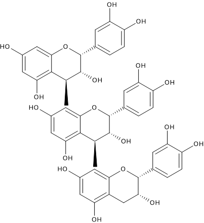
  2. Gu L., Kelm M.A., Hammerstone J.F., Beecher G., Holden J., Haytowitz D., Gebhardt S., and Prior R.L. Concentrations of proanthocyanidins in common foods and estimations of normal consumption. J Nutr 2004;134(3):613-617. doi:10.1093/jn/134.3.613
  3. Han X., Shen T. and Lou H. Dietary polyphenols and their biological significance. Int J Mol Sci 2007;9:950-988. doi:10.3390/i8090950
  4. Manach C., Scalbert A., Morand C., Rémésy C., and Jime´nez L. Polyphenols: food sources and bioavailability. Am J Clin Nutr 2004;79(5):727-747. doi:10.1093/ajcn/79.5.727
  5. Nandakumar V., Singh T., and Katiyar S.K. Multi-targeted prevention and therapy of cancer by proanthocyanidins. Cancer Lett 2008;269(2):378-387. doi:10.1016/j.canlet.2008.03.049
  6. Ottaviani J.I., Kwik-Uribe C., Keen C.L., and Schroeter H. Intake of dietary procyanidins does not contribute to the pool of circulating flavanols in humans. Am J Clin Nutr 2012;95:851-858. doi:10.3945/ajcn.111.028340
  7. Santos-Buelga C. and Scalbert A. Proanthocyanidins and tannin-like compounds: nature, occurrence, dietary intake and effects on nutrition and health. J Sci Food Agr 2000;80(7):1094-1117. doi:10.1002/(SICI)1097-0010(20000515)80:7<1094::AID-JSFA569>3.0.CO;2-1
  8. Tsao R. Chemistry and biochemistry of dietary polyphenols. Nutrients 2010;2:1231-1246. doi:10.3390/nu2121231
  9. Wang Y.,Chung S., Song W.O., and Chun O.K. Estimation of daily proanthocyanidin intake and major food sources in the U.S. diet. J Nutr 2011;141(3):447-452. doi:10.3945/jn.110.133900

Flavonols: structure, food sources, and benefits

Flavonols are polyphenols belonging to the flavonoid family.
They are colorless molecules that accumulate mainly in the outer and aerial tissues, therefore skin and leaves, of fruit and vegetables, since their biosynthesis is stimulated by light, whereas they are virtually absent in the flesh.
Flavonols are the most common flavonoids in fruit and vegetables, where they are generally present in relatively low concentrations.
Due to their widespread in nature and human diet, they should be taken into consideration when the positive effect on health associated with fruit and vegetable consumption is examined. Their effect is probably related to their ability to:

  • act as antioxidants;
  • act as anti-inflammatory agents;
  • act as anticancer factors;
  • regulate different cellular signaling pathways; an example is the action of quercetin, the most widespread flavonols, on the oxidative stress-induced MAPK activities.

Contents

Chemical structure

Chemically, these molecules differ from many other flavonoids since they have a double bond between positions 2 and 3 and an oxygen (a ketone group) in position 4 of the C ring, like flavones from which, however, they differ in the presence of a hydroxyl group at the position 3. Therefore, flavonol skeleton is a 3-hydroxyflavone.

3-Hydroxyflavone, the basic skeleton structure of flavonols
3-Hydroxyflavone

The 3-hydroxyl group can link a sugar, that is, it can be glycosylated.
Like many other flavonoids, most of them is found in fruit and vegetables, and in plant-derived foods, in glycosylated form. The sugar associated with flavonols is often glucose or rhamnose, but other sugars may also be involved, such as:

  • galactose;
  • arabinose;
  • xylose;
  • glucuronic acid.

Flavonols are mainly represented by glycosides of:

  • quercetin;
  • kaempferol;
  • myricetin;
  • isorhamnetin.
Skeletal formulas of flavonols such as quercetin and kaempferol
Flavonols

The most ubiquitous compounds are glycosylated derivatives of quercetin and kaempferol; in nature, these two molecules have respectively about 280 and 350 different glycosidic combinations.
Finally, it should be underlined that sugar moiety influences flavonol bioavailability.

Food sources

The major sources in human diet are:

  • fruit;
  • vegetables;
  • beverages such as red wine and tea.

In human diet, the richest source are capers, which contain up to 490 mg/100 g fresh weight (FW), but they are also abundant in onions, leeks, broccoli, curly kale, berries, for example blueberries, grapes and some herbs and spices, for example dill weed (Anethum graveolens). In these sources, their content ranges between 10 and 100 mg/100 g FW.
Even cocoa, green teablack tea, and red wine are good sources of flavonols. In wine, together with other polyphenols such as catechins, proanthocyanidins and low molecular weight polyphenols, they contribute to the astringency of the beverage.

Main flavonols in foods

The main flavonols in foods, listed in decreasing order of abundance, are quercetin, kaempferol, myricetin and ishoramnetin.

Quercetin

The richest sources of quercetin are capers, followed by onions, asparagus, lettuce and berries; in many other fruit and vegetables, it is present in smaller amounts, between 0.1 and 5 mg/100 g FW.
This flavonol is also present in cocoa and it could be one of its main protective agents against LDL oxidation.
Together with isoflavones, quercetin glycosides are the most well-absorbed polyphenols, followed by flavanones and catechins. Convesely, gallic acid derivatives of catechins are among the least well absorbed polyphenols, together with anthocyanins and proanthocyanidins.

Kaempferol

Typical dietary sources of kaempferol include vegetables, such as spinach, kale and endive, with concentrations between 0.1 and 27 mg/100 g FW, and some spices such as chives, fennel and tarragon, with concentrations between 6.5 and 19 mg/100 g FW.
Fruit is a poor source of the molecule, with content down to 0.1 mg/100 g FW.

Myricetin

Myricetin is the third most abundant flavonol. It is found in some spices, such as oregano, parsley, and fennel, with concentrations between 2 and 20 mg/100 g FW, but also in tea, 0.5-1.6 mg/100 ml, and red wine, 0-9.7 mg/100 ml.
In fruit, it is only found in high concentrations in berries, while in most other fruit and vegetables it is present in a content of less than 0.2 mg/100 g FW.

Isorhamnetin

A fourth flavonol, less abundant than the previous ones, is isorhamnetin. It is only present in some foods such as some spices: chives, 5.0-8.5 mg/100 g FW, fennel, 9.3 mg/100 g FW, tarragon, 5 mg/100 g FW.
In fruit and vegetables it is only present in almonds, with a concentration between 1.2 and 10.3 mg/100 g FW, pears and onions.

References

  1. de la Rosa L.A., Alvarez-Parrilla E., Gonzàlez-Aguilar G.A. Fruit and vegetable phytochemicals: chemistry, nutritional value, and stability. 1th Edition. Wiley J. & Sons, Inc., Publication, 2010
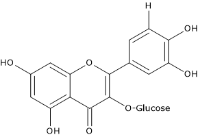
  2. Han X., Shen T. and Lou H. Dietary polyphenols and their biological significance. Int J Mol Sci 2007;9:950-988. doi:10.3390/i8090950
  3. Manach C., Scalbert A., Morand C., Rémésy C., and Jime´nez L. Polyphenols: food sources and bioavailability. Am J Clin Nutr 2004;79(5):727-747. doi:10.1093/ajcn/79.5.727
  4. Tsao R. Chemistry and biochemistry of dietary polyphenols. Nutrients 2010;2:1231-1246. doi:10.3390/nu2121231

Anthocyanins: structure, function, and health benefits

Anthocyanins are a subgroup of flavonoids, therefore they are polyphenols, which give plants their distinctive colors.
They are water soluble pigments and are present in the vacuolar sap of the epidermal tissues of flowers and fruit.
They are responsible for the colors of the most of the petals, fruits and vegetables, and of some varieties of cereals such as black rice. In fact, they impart red, pink and purple to blue colors to berries, red apples, red grapes, cherries, and of many other fruits, red lettuce, red cabbage, onions or eggplant, but also red wine.
Together with carotenoids, they are responsible for autumn leaf color and used commercially as food colours.
Finally, anthocyanins contribute to attract animals when a fruit is ready to eat or a flower is ready for pollination.

They are bioactive compounds found in plant foods that have a double interest for man:

  • the first one, a technological interest, due to their effects on the organoleptic characteristics of food products;
  • the other due to their healthy properties, being implicated in the protection against cardiovascular risk.
    In fact:

in vitro, they have an antioxidant activity, due to their ability to delocalize electrons and form resonance structures, and a protective role against oxidation of low density lipoproteins (LDL);

like other polyphenols, such as catechins, proanthocyanidins and other uncolored flavonoids, they can regulate different signaling pathways involved in cell growth, differentiation and survival.

Contents

Chemical structure

The basic chemical structure is flavylium cation (2-phenyl-1-benzopyrilium), which links hydroxyl (-OH) and/or methoxyl (-OCH3) groups, and one or more sugars.
The sugar-free molecule is called anthocyanidins.

Skeletal formula of the basic skeleton of anthocyanins
Flavylium Cation

Depending on the number and position of hydroxyl and methoxyl groups, various anthocyanidins have been described, and of these, six are commonly found in vegetables and fruits:

  • pelargonidin
  • cyaniding
  • delphinidin
  • petunidin
  • peonidin
  • malvidin
Skeletal formulas of different types of anthocyanins
Antocyanins

Anthocyanins, as most of the other flavonoids, are present in plants and plant foods in the form of glycosides, that is, linked to one or more sugar units.
The most common carbohydrates present in these natural pigments are:

The sugars are linked mainly to the C3 position as 3-monoglycosides, to the C3 and C5 positions as diglycosides, with the possible forms: 3-diglycosides, 3,5-diglycosides, and 3-diglycoside-5-monoglycosides. Glycosylations have been also found at C7, C3′ and C5′ positions.
The structure of these molecules is further complicated by the bond to the sugar unit of different acyl substituents such as:

  • aliphatic acids, such as acetic, malic, succinic and malonic acid;
  • cinnamic acids (aromatic substituents), such as sinapic, ferulic and p-coumaric acid;
  • finally, there are pigments with both aromatic and aliphatic substituents.

Furthermore, some anthocyanins have several acylated sugars in the molecule; these anthocyanins are sometimes called polyglycosides.

Depending on the type of hydroxylation, methoxylation and glycosylation patterns, and the different substituents linked to the sugar units, more than 500 different anthocyanins have been identified that are based on 31 anthocyanidins. Among these 31 monomers:

  • 30 percent are from cyanidin;
  • 22 percent are from delphinidin;
  • 18 percent are from pelargonidin.

Methylated derivatives of cyanidin, delphinidin and pelargonidin, namely peonidin, malvidin, and petunidin, all together represent 20 percent of the anthocyanins.
Therefore, up to 90 percent of the most frequently encountered anthocyanins are related to delphinidin, pelargonidin, cyanidin, and their methylated derivatives.

Role of pH

The color of these molecules is influenced by the pH of the vacuole where they are stored, ranging in color from:

  • red, under very acidic conditions;
  • to purple-blue, in intermediate pH conditions;
  • until yellow-green, in alkaline conditions.

In addition to the pH, the color of these flavonoids can be affected by the degree of hydroxylation or methylation pattern of the A and B rings, and by glycosylation pattern.
Finally, the color of certain plant pigments result from complexes between anthocyanins, flavones and metal ions.
It should be noted that anthocyanins are often used as pH indicators thanks to the differences in chemical structure that occur in response to changes in pH.

References

  1. de la Rosa L.A., Alvarez-Parrilla E., Gonzàlez-Aguilar G.A. Fruit and vegetable phytochemicals: chemistry, nutritional value, and stability. 1th Edition. Wiley J. & Sons, Inc., Publication, 2010
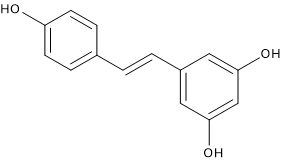
  2. de Pascual-Teresa S., Moreno D.A. and García-Viguera C. Flavanols and anthocyanins in cardiovascular health: a review of current evidence. Int J Mol Sci 2010;11:1679-1703. doi:10.3390/ijms11041679
  3. Escribano-Bailòn M.T., Santos-Buelga C., Rivas-Gonzalo J.C. Anthocyanins in cereals. J Chromatogr A 2004:1054;129-141. doi:10.1016/j.chroma.2004.08.152
  4. Han X., Shen T. and Lou H. Dietary polyphenols and their biological significance. Int J Mol Sci 2007;9:950-988. doi:10.3390/i8090950
  5. Manach C., Scalbert A., Morand C., Rémésy C., and Jime´nez L. Polyphenols: food sources and bioavailability. Am J Clin Nutr 2004;79(5):727-747. doi:10.1093/ajcn/79.5.727
  6. Ottaviani J.I., Kwik-Uribe C., Keen C.L., and Schroeter H. Intake of dietary procyanidins does not contribute to the pool of circulating flavanols in humans. Am J Clin Nutr 2012;95:851-858. doi:10.3945/​ajcn.111.028340
  7. Tsao R. Chemistry and biochemistry of dietary polyphenols. Nutrients 2010;2:1231-1246. doi:10.3390/nu2121231

Proanthocyanidins: structure and intestinal absorption

Proanthocyanidins or condensed tannins, also called pycnogenols and leukocyanidins, are polyphenolic compounds, in particular they are a flavonoid subgroup, widely distributed in the plant kingdom, second only to lignin as the most abundant phenol in nature.
They are present in high concentrations in various parts of the plants such as flowers, fruits, berries, seeds, for example in grape seeds, and bark, like pine bark.
Together with anthocyanins and their oxidation products, and catechins, they are the most abundant flavonoids in human diet and it has been suggested that they constitute a significant fraction of the polyphenols ingested in the Western diet.
Therefore, condensed tannins should be taken into consideration when the epidemiological association between the intake of polyphenols, especially flavonoids, and chronic diseases are examined.

Contents

Chemical structure

Condensed tannins have a complex chemical structure being oligomers, from dimers to pentamers, or polymers, from six or more units up to 60, of catechins or flavanols, joined by carbon-carbon bonds.

Basic skeleton structure of procyanidins, a type of proanthocyanidins
Basic Skeleton of Procyanidins

They may consist exclusively of:

  • (epi)catechin, and they are named procyanidins;
  • (epi)afzelechin, and they are named propelargonidins;
  • (epi)gallocatechin, and they are named prodelphinidins.

Propelargonidins and prodelphinidins are less common in nature and in foods than procyanidins.

Depending on the bonds between monomers, proanthocyanidins have a:

  • B-type structure, if the polymerization occurs via carbon-carbon bond between the position 8 of the terminal unit and the 4 of the extender (or C4-C6);
  • A-type structure, less frequent, if monomers are doubly linked via an ether bond C2-O-C7 or C2-O-C5 plus a B-type bond.

Procyanidins

The most common dimers are B-type procyanidins, B1 to B8, formed by catechin or epicatechin; in B1, B2, B3 and B-4 dimers, the two flavanol units are joined by a C4-C8 bond; in B5, B6, B7 and B8 dimers the two units are joined by C4-C6 bond.

Skeletal formulas of procyanidin B1, B2, B3, and B4
Procyanidins B1, B2, B3, and B4

Procyanidin C1 is a B-type trimer.

Procyanidin A-2 is an example of A-type procyanidin.

Intestinal absorption

Condensed tannins are poorly absorbed from the intestine; together with anthocyanins and gallic acid ester derivatives of tea catechins, they are the least well-absorbed polyphenols.
It seems that low molecular weight oligomers (2-3 monomers) may be absorbed as such while polymers are not.
In the systemic circulation, dimers reach concentrations of two orders of magnitude lower than those of catechins.
It seems that condensed tannins with a degree of polymerization greater than three transit into the stomach and small intestine without significant modifications, and then, into the large intestine, they are catabolized by gut microbiota, which is part of the human microbiota, with production of phenylpropionic, phenilvaleric and phenylacetic acids. These degradation products have been suggested to be the major metabolites of proanthocyanidins in healthy humans.

Procyanidins and catechins

It had been proposed that the catabolism of procyanidins in the gastrointestinal tract lead to the release of monomeric catechins, thus indirectly contributing to their systemic pool in humans. In recent years, it has been shown that this does not happen because procyanidins do not significantly contribute to:

  • the concentration of catechin metabolites in the systemic circulation;
  • the total catechin metabolites excreted in the urine;
  • finally, they do not significantly affect plasma metabolite profile derived from catechol-O-methyltransferase activity.

Therefore, analyzing the potential health benefits associated with the intake of foods containing these phytochemicals, catechins and procyanidins should be considered distinct classes of related compounds.

References

  1. de la Rosa L.A., Alvarez-Parrilla E., Gonzàlez-Aguilar G.A. Fruit and vegetable phytochemicals: chemistry, nutritional value, and stability. 1th Edition. Wiley J. & Sons, Inc., Publication, 2010
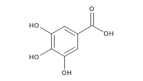
  2. Gu L., Kelm M.A., Hammerstone J.F., Beecher G., Holden J., Haytowitz D., Gebhardt S., and Prior R.L. Concentrations of proanthocyanidins in common foods and estimations of normal consumption. J Nutr 2004;134(3):613-617. doi:10.1093/jn/134.3.613
  3. Han X., Shen T. and Lou H. Dietary polyphenols and their biological significance. Int J Mol Sci 2007;9:950-988. doi:10.3390/i8090950
  4. Manach C., Scalbert A., Morand C., Rémésy C., and Jime´nez L. Polyphenols: food sources and bioavailability. Am J Clin Nutr 2004;79(5):727-747. doi:10.1093/ajcn/79.5.727
  5. Nandakumar V., Singh T., and Katiyar S.K. Multi-targeted prevention and therapy of cancer by proanthocyanidins. Cancer Lett 2008;269(2):378-387. doi:10.1016/j.canlet.2008.03.049
  6. Ottaviani J.I., Kwik-Uribe C., Keen C.L., and Schroeter H. Intake of dietary procyanidins does not contribute to the pool of circulating flavanols in humans. Am J Clin Nutr 2012;95:851-858. doi:10.3945/ajcn.111.028340
  7. Santos-Buelga C. and Scalbert A. Proanthocyanidins and tannin-like compounds: nature, occurrence, dietary intake and effects on nutrition and health. J Sci Food Agr 2000;80(7):1094-1117. doi:10.1002/(SICI)1097-0010(20000515)80:7<1094::AID-JSFA569>3.0.CO;2-1
  8. Tsao R. Chemistry and biochemistry of dietary polyphenols. Nutrients 2010;2:1231-1246. doi:10.3390/nu2121231
  9. Wang Y.,Chung S., Song W.O., and Chun O.K. Estimation of daily proanthocyanidin intake and major food sources in the U.S. diet. J Nutr 2011;141(3):447-452. doi:10.3945/jn.110.133900

Catechins: structure and food sources

Catechins or flavanols, with flavonols such as quercetin, and flavones such as luteolin, are a subgroup of flavonoids among the most widespread in nature.
Flavanols and proanthocyanidins, together with anthocyanins and their oxidation products, are the most abundant flavonoids in human diet.

Contents

Chemical structure

Chemically they differ from many other flavonoids as:

  • they lack the double bond between positions 2 and 3 of the C ring;
  • they not have a keto group at position 4;
  • they have a hydroxyl group in position 3, and for this reason they are also called flavan-3-ols.
Basic skeleton structure of catechins
Basic Skeleton of Catechins

Another distinctive feature of flavan-3-ols is their ability to form oligomers, consisting of two to ten units, or polymers, consisting of from eleven or more units up to 60 units, called proanthocyanidins or condensed tannins.

Food sources

Flavanols commonly found in plant-derived food products are catechin, epicatechin, gallocatechin, epigallocatechin, and their gallic acid ester derivatives: catechin gallate, gallocatechin gallate, epicatechin gallate, and epigallocatechin gallate or EGCG.

Skeletal formulas of catechin, epicatechin, gallocatechin, epigallocatechin
Catechins

Flavanols present with higher frequency are catechin and epicatechin, which are also among the most common known flavonoids, and almost as popular as the related flavonol quercetin.
Cocoa and green tea are by far the richest sources in flavanols. In these foods the main flavonoids are catechin and epicatechin, but also their gallic acid ester derivatives, the gallocatechins. Cocoa is also a good source of epigallocatechin.

Structural formulas of gallic acid ester derivatives of catechins
Gallic Acid Ester Derivatives of Catechins

However, they are also present in many fruits, especially in the skins of apples, blueberries (Vaccinium myrtillus) and grapes, in vegetables, red wine and beer, and peanuts.
As in many cases flavanols are present in the seeds or peels of fruits and vegetables, their intake may be limited by the fact that these parts are discarded during processing or while eaten.
Furthermore, in contrast to other flavonoids, catechins are not glycosylated in foods.
Proanthocyanidins, that is polymeric flavan-3-ols, are also commonly found in plant-derived food products. Their presence has been reported in the skin of peanuts and almonds, as in the berries.

Green and black tea

Green tea is an excellent source of flavonoids. The main flavonoids present in the leaves of the tea, like in cocoa beans, are catechin and epicatechin, monomeric flavanols, together with their gallate derivatives such as EGCG.
Epigallocatechin gallate is the most abundant catechin in green tea and it seems to have an important role in determining green tea benefits, as the reduction of:

  • vascular inflammation;
  • blood pressure;
  • concentration of oxidized LDLs.

Black tea, which is a fermented tea, contains fewer monomeric flavanols, as they are oxidized during fermentation of the leaves to more complex polyphenols such as theaflavins, specifically theaflavin digallate, theaflavin-3-gallate, and theaflavin-3′-gallate, which are all dimers, and thearubigins, which are polymers.
Theaflavins and thearubigins are present only in the tea. Their concentrations in brewed tea are between 50- and 100-folds lesser than in tea leaves.
It should be noted that tea epicatechins are remarkably stable to heat in acidic environment: at pH 5, only about 15% is degraded after seven hours in boiling water (therefore, adding lemon juice to brewed tea does not cause any reduction in their content).

Cocoa and cocoa products

Cocoa has the highest content of polyphenols and flavanols per serving, a concentration greater than those found in green tea and red wine. Most of the flavonoids present in cocoa beans and derived products, such as black chocolate, are catechin and epicatechin, monomeric flavanols, but also epigallocatechin, and their derivatives such as the gallocatechins; among polymers, proanthocyanidins are also important.

Fruits, vegetables, and legumes

Catechin and epicatechin are the main flavanols in fruits. They are found in many fruits in different concentrations, respectively, between 5-3 and 0.5-6 mg/100 g fresh weight.
On the contrary, gallocatechin, epicatechin gallate, epigallocatechin, and epigallocatechin gallate are present in various fruits such as red grapes, berries, apples, peaches and plums, but in very low concentrations, less than 1mg/100 g fresh weight.
Except for lentils and broad beans, few legumes and vegetables contain catechins, and in very low concentrations, less than 1.5 mg/100 g fresh weight.

References

  1. de la Rosa L.A., Alvarez-Parrilla E., Gonzàlez-Aguilar G.A. Fruit and vegetable phytochemicals: chemistry, nutritional value, and stability. 1th Edition. Wiley J. & Sons, Inc., Publication, 2010
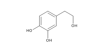
  2. de Pascual-Teresa S., Moreno D.A. and García-Viguera C. Flavanols and anthocyanins in cardiovascular health: a review of current evidence. Int J Mol Sci 2010;11:1679-1703. doi:10.3390/ijms11041679
  3. Han X., Shen T. and Lou H. Dietary polyphenols and their biological significance. Int J Mol Sci 2007;9:950-988. doi:10.3390/i8090950
  4. Manach C., Scalbert A., Morand C., Rémésy C., and Jime´nez L. Polyphenols: food sources and bioavailability. Am J Clin Nutr 2004;79(5):727-747. doi:10.1093/ajcn/79.5.727
  5. Tsao R. Chemistry and biochemistry of dietary polyphenols. Nutrients 2010;2:1231-1246. doi:10.3390/nu2121231

Flavonoids: definition, structure, and classification

Flavonoids are the most abundant polyphenols in human diet, representing about 2/3 of all those ones ingested. Like other phytochemicals, they are the products of secondary metabolism of plants and, currently, it is not possible to determine precisely their number, even if over 4000 have been identified.
In fruits and vegetables, they are usually found in the form of glycosides and sometimes as acylglycosides, while acylated, methylated and sulfate molecules are less frequent and in lower concentrations.
They are water-soluble and accumulate in cell vacuoles.

Contents

Chemical structure

Their basic structure is a skeleton of diphenylpropane, i.e., two benzene rings linked by a three carbon chain that forms a pyran ring, namely, a heterocyclic ring containing oxygen, closed with the benzene ring A, which is called ring C. Therefore, their structure is also referred to as C6-C3-C6.

Basic skeleton structure of flavonoids, the most abundant polyphenols in human diet
Basic Skeleton of Flavonoids

In most cases, B ring is attached to position 2 of C ring, but it can also bind in position 3 or 4. This, together with the structural features of the ring B and the patterns of glycosylation and hydroxylation of the three rings, makes the flavonoids one of the larger and more diversified groups of phytochemicals, so not only of polyphenols, in nature.
Their biological activities, for example they are potent antioxidants, depend both on the structural characteristics and the pattern of glycosylation.

Classification

They can be subdivided into different subclasses depending on the carbon of the C ring on which B ring is attached, and the degree of unsaturation and oxidation of the C ring.
Flavonoids in which B ring is linked in position 3 of the ring C are called isoflavones; those in which B ring is linked in position 4, neoflavonoids, while those in which the B ring is linked in position 2 can be further subdivided into several subgroups on the basis of the structural features of the C ring. These subgroup are: flavones, flavonols, flavanones, flavanonols, flavanols or catechins and anthocyanins.
Finally, flavonoids with open C ring are called chalcones.

Basic skeleton structure of flavonoid subclasses
Flavonoid Subclasses

Flavones

They have a double bond between positions 2 and 3 and a ketone in position 4 of the C ring. Most flavones of vegetables and fruits has a hydroxyl group in position 5 of the A ring, while the hydroxylation in other positions, for the most part in position 7 of the A ring or 3′ and 4′ of the B ring may vary according to the taxonomic classification of the particular vegetable or fruit.
Glycosylation occurs primarily on position 5 and 7, methylation and acylation on the hydroxyl groups of the B ring.
Some flavones, such as nobiletin and tangeretin, are polymethoxylated.

Flavonols

Compared to flavones, they have a hydroxyl group in position 3 of the C ring, which may also be glycosylated. Again, like flavones, flavonols are very diverse in methylation and hydroxylation patterns as well, and, considering the different glycosylation patterns, they are perhaps the most common and largest subgroup of flavonoids in fruits and vegetables. For example, quercetin is present in many plant foods.

Flavanones

Flavanones, also called dihydroflavones, have the C ring saturated; therefore, unlike flavones, the double bond between positions 2 and 3 is saturated and this is the only structural difference between the two subgroups of flavonoids.
The flavanones can be multi-hydroxylated, and several hydroxyl groups can be glycosylated and/or methylated.
Some have unique patterns of substitution, for example, furanoflavanones, prenylated flavanones, pyranoflavanones or benzylated flavanones, giving a great number of substituted derivatives.
Over the past 15 years, the number of flavanones discovered is significantly increased.

Flavanonols

Flavanonols, also called dihydroflavonols, are the 3-hydroxy derivatives of flavanones; they are an highly diversified and multisubstituted subgroup.

Isoflavones

As anticipated, isoflavones are a subgroup of flavonoids in which the B ring is attached to position 3 of the C ring. They have structural similarities to estrogens, such as estradiol, and for this reason they are also called phytoestrogens.

Catechins

Catechins are also referred to flavan-3-ols as the hydroxyl group is almost always bound to position 3 of C ring; they are called flavanols as well.
They have two chirality centers in the molecule, on positions 2 and 3, then four possible diastereoisomers. Epicatechin is the isomer with the cis configuration and catechin is the one with the trans configuration. Each of these configurations has two stereoisomers, namely, (+)-epicatechin and (-)-epicatechin, (+)-catechin and (-)-catechin.
(+)-Catechin and (-)-epicatechin are the two isomers most often present in edible plants.
Another important feature of flavanols, particularly of catechin and epicatechin, is the ability to form polymers, called proanthocyanidins or condensed tannins. The name “proanthocyanidins” is due to the fact that an acid-catalyzed cleavage produces anthocyanidins.
Proanthocyanidins typically contain 2 to 60 monomers of flavanols.
Monomeric and oligomeric flavanols, consisting of 2 to 7 monomers, are strong antioxidants.

Anthocyanidins

Chemically, anthocyanidins are flavylium cations and are generally present as chloride salts.
They are the only colored flavonoids, and, with carotenoids, impart color to plants. Moreover, they are used commercially as food colours.
Anthocyanins are glycosides of anthocyanidins. Sugar units are bound mostly to position 3 of the C ring and they are often conjugated with phenolic acids, such as ferulic acid.
The color of the anthocyanins depends on the pH and also by methylation or acylation at the hydroxyl groups on the A and B rings.

Chalcones

Chalcones and dihydrochalcones are flavonoids with open structure; they are classified as flavonoids because they have similar synthetic pathways.

References

  1. de la Rosa L.A., Alvarez-Parrilla E., Gonzàlez-Aguilar G.A. Fruit and vegetable phytochemicals: chemistry, nutritional value, and stability. 1th Edition. Wiley J. & Sons, Inc., Publication, 2010
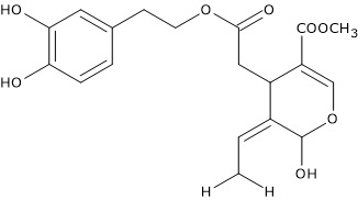
  2. Han X., Shen T. and Lou H. Dietary polyphenols and their biological significance. Int J Mol Sci 2007;9:950-988. doi:10.3390/i8090950
  3. Manach C., Scalbert A., Morand C., Rémésy C., and Jime´nez L. Polyphenols: food sources and bioavailability. Am J Clin Nutr 2004;79(5):727-747. doi:10.1093/ajcn/79.5.727
  4. Panche A.N., Diwan A.D., and Chandra S.R. Flavonoids: an overview. J Nutr Sci. 2016;5:e47. doi:10.1017/jns.2016.41
  5. Tsao R. Chemistry and biochemistry of dietary polyphenols. Nutrients 2010;2:1231-1246. doi:10.3390/nu2121231

Polyphenols: structure, classification, and food sources

Polyphenols are among the most important, and certainly the most numerous, phytochemicals present in the plant kingdom.
Currently, over 8,000 phenolic structures have been identified, of which more than 4,000 belonging to the class of flavonoids, and several hundred occur in edible plants.
However, it is thought that the total content of polyphenols in plants is underestimated as many of the phenolic compounds present in fruits, vegetables and derivatives have not yet been identified, escaping the methods and techniques of analysis used, and the composition in polyphenols for most fruits and some varieties of cereals is not yet known.
They are present in many edible plants, both for men and animals, and it is thought to be their presence, along with that of other molecules such as carotenoids, vitamin C or vitamin E, the responsible for the healthy effects of fruits and vegetables.
In the human diet, they are the most abundant natural antioxidants, and the main sources are fruits, vegetables, whole grains, but also other types of foods and beverages derived from them, such as red wine, rich in resveratrol, the extra virgin olive oil, rich in hydroxytyrosol, chocolate or tea, in particularly green tea, rich in epigallocatechin gallate or EGCG.

Contents

Chemical structure

The term polyphenols refers to a wide variety of molecules that can be divided into many subclasses, subdivisions that can be made on the basis of their origin, biological function, or chemical structure.
Chemically, they are compounds with structural phenolic features, which can be associated with different organic acids and carbohydrates.

Model of phenol, the basic structural feature of polyphenols
Ball-and-Stick Model of Phenol

In plants, the most part of them are linked to sugars, and therefore they are in the form of glycosides. Carbohydrates and organic acids can be bound in different positions on polyphenol skeletons.
Among polyphenols, there are simple molecules, such as phenolic acids, or complex structures such as proanthocyanidins, that are highly polymerized molecules.

Classification

They can be classified into different classes, according to the number of phenolic rings in their structure, the structural elements that bind these rings each others, and the substituents linked to the rings. Therefore, two main groups can then be identified: the flavonoid group and the non-flavonoid group.
Flavonoids share a structure formed by two aromatic rings, indicated as A and B, linked together by three carbon atoms forming an oxygenated heterocycle, the C ring. They can be further subdivided into six main subclasses, as a function of the type of heterocycle, the C ring, that is involved:

Non-flavonoids can be subdivided into:

  • simple phenols;
  • phenolic acids;
  • benzoic aldehydes;
  • hydrolyzable tannins;
  • acetophenones and phenylacetic acids;
  • hydroxycinnamic acids;
  • coumarins;
  • benzophenones;
  • xanthones;
  • stilbenes;
  • lignans;
  • secoiridoids.

Variability of polyphenol content of plants and plant products

Although several classes of phenolic molecules, such as quercetin, a flavonol, are present in most plant foods, such as tea, wine, cereals, legumes, fruits, fruit juices, etc., other classes are found only in a particular type of food, for example flavanones in citrus, isoflavones in soya, phloridzin in apples, etc.
However, it is common that different types of polyphenols are in the same product; for example, apples contain flavanols, chlorogenic acid, hydroxycinnamic acids, glycosides of phloretin, glycosides of quercetin and anthocyanins.
The polyphenol composition may also be influenced by other parameters such as environmental factors, the degree of ripeness at harvest time, household or industrial processing, storage, and plant variety. From currently available data, it seems that the fruits with the highest content of polyphenols are strawberries, lychees and grapes, and the vegetables are artichokes, parsley and brussels sprouts. Melons and avocados have the lowest concentrations.

References

  1. de la Rosa L.A., Alvarez-Parrilla E., Gonzàlez-Aguilar G.A. Fruit and vegetable phytochemicals: chemistry, nutritional value, and stability. 1th Edition. Wiley J. & Sons, Inc., Publication, 2010
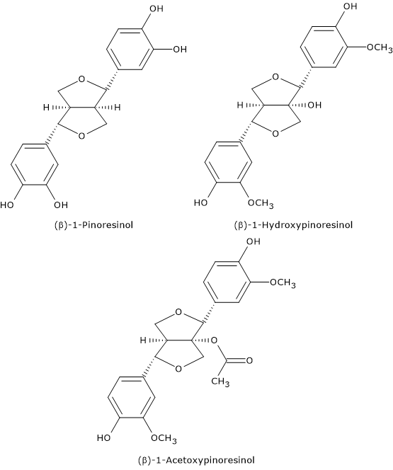
  2. Han X., Shen T. and Lou H. Dietary polyphenols and their biological significance. Int J Mol Sci 2007;9:950-988. doi:10.3390/i8090950
  3. Manach C., Scalbert A., Morand C., Rémésy C., and Jime´nez L. Polyphenols: food sources and bioavailability. Am J Clin Nutr 2004;79(5):727-747. doi:10.1093/ajcn/79.5.727
  4. Tsao R. Chemistry and biochemistry of dietary polyphenols. Nutrients 2010;2:1231-1246. doi:10.3390/nu2121231